Medically reviewed and approved by a board-certified member
Zoology
ULTRA STRUCTURE OF MUSCLE
By BS MediaTwitter Profile | Updated: Thursday, 25 May 2017 19:51 UTC
The muscles of a fully developed adult male can produce sufficient power to lift a weight of about 25 tonnes.
- In male, all the muscles make up 42% of the body weight. In woman, the muscles make up 36% of the body weight.
- The body consists of 650 muscles. 620 muscles work together to move 206 bones of the skeleton.
- Remaining 30 muscles are needed to ensure the passage of food through alimentary canal, to circulate the flood round the body and operate certain internal organs.
- The muscles by themselves donot contract.
- They are under the controle of central nervous system [CNS].
- For example, if we wish to pick up an object, our CNS must know the tension, length and position of the arm muscles inorder to cause the correct amount of contraction to move the arm.
- The CNS must stop the contractions when the arm reaches the object, maintain sufficient contraction to hold the object. Then CNS cause the muscle contractions required to move the arm away.
- The muscles are biological machines which convert chemical energy into force and mechanical work.
- The energy for contraction, as for other life activities, comes from the chemical reaction between the food stuffs that we eat and the oxygen that we breath.
- Muscles therefore need a good blood supply to bring essential food and oxygen.
- The main property of the muscle is contraction.
- The muscles contract due to mechanical, thermal, chemical and electrical stimuli.
- The muscles receive stimuli through motor nerves.
- Before studying the muscle contraction, it is better to learn about the structure of a muscle.
MUSCLES AND ITS STRUCTURAL ORGANISATION
- For the movement of the body and its parts in an animal, muscular tissue is present.
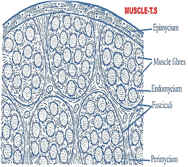
- The muscular tissue shows myocytes or muscle fibres. Individual muscle fibre is surrounded by fine fibrous • connective tissue called endomycium.
- The parrallel bundles of muscle fibres are called fasciculi or fascicles.
- Each fasciculus is surrounded by perimycium. The entire muscle is usually wrapped with a fibrous connective tissue called epimycium.
- The cell wall or plasma membrane of muscle fibre is called "sarcolemma".
- The cytoplasm is called "sarcoplasm", which consists of many nuclei nearer to the sarcolemma.
- Hence muscle cells or muscle fibres are called "syncytial" cells.
- The sarcoplasm also consists of many, longitudinally arranged fibrils called "myofibrils".
- These are the contractile structures. The details of myofibrils can be seen only under the electron microscope.
MUSCLE - ULTRA STRUCTURE
The ultra-structure of muscle is well known by the structure of longitudinaly arranged fibrils called myofibrils.
MYO FIBRIL
- Each myofibril consists of "light" and "dark" bands.
- The light band is called "isotropic" or "I-band". The dark band is called "anisotropic" or "A-band".
- These bands are arranged alternately.
- The bands give a striated appearance to the myofibrils.
- Hence the muscles are called "striatedmuscles".
- The bands are made up of filaments, which inturn are made up of muscle proteins.
"Themuscle proteins are Actin and Myosin".
- The light T-band is made up of thin Actin filaments.
- Whereas the Dark 'A'-band is made up of thick myosin filaments. The atomic weight of actin atom is 70,000 A.M.U.
- Hence the l-band is light. The atomic weight of myosin atom is 4,50,000 A.M.U. Hence the 'A'-band is dark.
- The protein actin normally exists with calcium ion. Whereas the protein myosin exists with magnesium ion.
- The light l-band is divided into two equal parts by black line called "Z" membrane or "krause's membrane".
- The region of myofibril between the two successive 'Z' membranes is' called "sarcomere".
- One full A-band and two halves of two l-bands, which lie on either sides of 'A' band.
- The sarcomeres are the fundamental units of myofibrils.
- The Dark 'A'- band is also divided into two equal parts by light membrane called "H" zone or "Henson's disd", which consists of black line at its centre, called 'M' line.
- Glutei, buttock muscle of Frog is taken and its contents like, 'A.T.P.' is extracted by pressing.
- When such muscle is kept in A.T.P. liquid, it contracts. The above experiment shows that Actin, myosin, A.T.P, and calcium ions are essential for muscle contraction. A.T.P. and CP are synthesized by the oxidation of the food materials.
- So the mitochondria play an important role for muscle contraction.
- Hence the mitochondria in muscle cell are called "sarcosomeres". The Ca++ and Mg++ are secreted by endoplasmic reticulum.
- Hence E.R in muscle cell is called sarcoplasmic reticulum.
Chemistry of Muscle
i. The muscle contains 75% of water and 25% of solids.
ii. Most of the solids are soluble proteins such as actin, myosin, mysinogen, paramysinogen.
iii. Non protein organic substances present in the muscles are ATP, phosphocreatine, creatine and urea.
iv. Important in the muscle is glycogen.
v. Mineral elements present in a muscle are sodium, potassium, calcium, phosphorus and magnesium, out of which potassium occurs abundantly.
vi. Muscles have their own oxygen - carrying iron - protein pigment myoglobin or muscle haemoglobin.
Three kinds of muscles are seen in animals.
1. striped muscle or striated,
2. unstriped or unstriated
3. Cardiac muscle.
- The cardiac muscles are striated, but they are involuntary.
- The actin and myosin proteins are arranged as bands like in striped muscles.
- The smooth muscles also have actin and myosin, which are not aggregated into filaments.
End of the article