a
b
c
d
e
f
g
h
i
j
k
l
m
n
o
p
q
r
s
t
u
v
w
x
y
z
#
Pathology / Clinical Pathology

7 Vital Elements for Ensuring Lab Excellence through Maintenance, Repair, and Calibration

Acetest Tablet Test for Ketone Bodies: Principle, Procedure, and Interpretation
Acid Phosphatase (AP): Total and Prostatic Acid Phosphatase
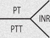
Activated Partial Thromboplastin Time (APTT), Partial Thromboplastin Time (PTT), Prothrombin Time (PT) and INR
Adrenal Gland Hormones and Interpretation
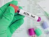
Adrenocorticotropic Hormone (ACTH)
Bilirubin: Total, Direct and Indirect Bilirubin (Different Types of Jaundice)

Biochemical Analysis of Semen for Investigation of Infertility
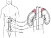
Biochemical Tests Used to Assess Renal Function
Biuret Test for Proteinuria: Principle, Procedure, and Interpretation

Blood Sample Preservation Method for PT/APTT Testing
Chemical Examination of Feces
Chemical Examination of Urine