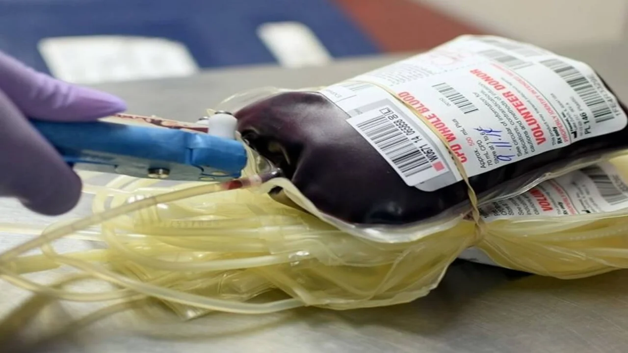
Preparation of Blood Components from Whole Blood for Transfusion
Discover the preparation methods for blood components, including packed red blood cells, platelet-rich plasma, leukocyte-poor red cells, washed red cells, frozen red cells, irradiated red cells, platelets, fresh frozen plasma, and cryoprecipitate.

A single donation of whole blood can be separated into multiple components, enabling treatment for more than one patient. From one unit of whole blood, three primary components can be derived: packed red blood cells, platelets, and fresh frozen plasma or cryoprecipitate. This approach minimizes waste by storing each component at an optimal temperature, facilitating specific replacement therapy and preventing the unnecessary transfusion of blood elements that a patient may not need.
Methods of Blood Collection for Component Preparation
There are two primary methods for collecting blood for the preparation of its components:
- Single Whole Blood Donation: The development of double and triple bags with integral closed tubing systems has greatly advanced the preparation of blood components. After a unit of whole blood is collected in a primary bag, the components are separated using differential centrifugation, which takes advantage of their varying densities. This method ensures that the different blood products are transferred from one bag to another while maintaining a closed circuit. By doing so, the sterility of the products is preserved. It is crucial to process the blood within six hours of collection to ensure optimal separation (Refer to Flowchart 1 and Figure 1).
- Apheresis: Apheresis is a more advanced procedure wherein the donor is connected to an automated cell separator machine, which acts as a centrifuge. Through this process, whole blood is withdrawn from the donor, the required component is isolated and retained, while the remaining blood is returned to the donor. Depending on which component is retained, the procedure can be referred to as plateletpheresis, leukapheresis, or plasmapheresis.
Transfusion and Blood Component Processing
When whole blood is collected, it can be processed into several components within six hours. Light centrifugation separates red cells from platelets and plasma. Following that, a more intense centrifugation of platelet-rich plasma results in the separation of platelets and plasma (Refer to Flowchart 1).
- Whole blood
- Light spin
- Packed red cells
- Leukocyte-poor red cells
- Washed red cells
- Frozen red cells
- Irradiated red cells
- Platelet-rich plasma
- Heavy spin
- Platelet concentrate
- Plasma
- Fresh frozen plasma
- Cryoprecipitate
- Heavy spin
- Packed red cells
- Light spin

Breakdown of blood components:
- Packed Red Cells: These are red blood cells with minimal plasma.
- Platelet-rich Plasma: Plasma that is rich in platelets.
- Leukocyte-poor Red Cells: Red blood cells with a reduced number of white blood cells.
- Washed Red Cells: Red blood cells free of plasma proteins and other contaminants.
- Frozen Red Cells: Red cells preserved with a cryoprotective agent.
- Irradiated Red Cells: Red cells treated with gamma rays to inactivate lymphocytes.
Terms in Blood Transfusion Therapy
Table 1 provides key definitions commonly used in blood transfusion therapy.
| Term | Definition |
|---|---|
| Blood product | A therapeutic substance prepared from human blood |
| Whole blood | One unit of non-separated donor blood collected in an appropriate container containing the anticoagulant-preservative solution |
| Blood component | A constituent separated from whole blood by differential centrifugation or that is obtained directly from the donor by apheresis |
| Plasma derivative | Human plasma proteins obtained from multiple donor units of plasma under pharmaceutical manufacturing conditions. These products are heat-treated or chemical-treated to inactivate lipid-enveloped viruses. |
| Plasma derivatives like factor concentrates and immunoglobulins can also be prepared by recombinant DNA technology. | |
Whole Blood
Whole blood refers to a unit of donor blood preserved in an anticoagulant-preservative solution, typically citrate phosphate dextrose adenine (CPDA-1). Each unit contains about 400 ml of blood, including 49 ml of anticoagulant. It consists of cellular components and plasma, but lacks functional platelets and certain labile coagulation factors like Factor V and Factor VIII. Whole blood is typically stored at temperatures between 4°C and 6°C and can be kept for up to 35 days. Transfusion of whole blood should begin within 30 minutes of its removal from cold storage and must be completed within four hours. Transfusion of one unit raises hemoglobin by 1 gm/dl or hematocrit by 3%.
Indications for Whole Blood Transfusion:
- Acute blood loss with hypovolemia.
- Neonatal exchange transfusions.
- When red cell concentrates are unavailable.
Whole blood transfusion is contraindicated in chronic anemia cases with compromised cardiovascular function (Refer to Table 2).
| Indications | Contraindications |
|---|---|
|
Chronic anemia with compromised cardiovascular function |
Blood Components
The blood components are categorized into cellular and plasma components (Refer to Table 3).
| Component Type | Examples |
|---|---|
| Cellular components |
|
| Plasma components |
|
Red Cell Components
1. Packed Red Cells
Packed red cells are prepared by removing most plasma from a unit of whole blood, yielding a concentrated volume of red cells with a hematocrit of 70-75%. These cells are separated either by allowing the blood to sediment in a refrigerator at 2-6°C or is spun in a refrigerated centrifuge. Supernatant plasma is then separated from red cells in a closed system by transferring it to the attached empty satellite bag. Red cells and a small amount of plasma are left behind in the primary blood bag. Packed red cells have a high viscosity and therefore the rate of infusion is slow. The transfusion of one unit of packed red cells typically increases hemoglobin by 1 gram per deciliter (or increases hematocrit by 3%).
Indications for Packed Red Cells Transfusion:
- Anemia: Chronic severe anemia, severe anemia with congestive cardiac failure, anemia in elderly.
- Acute blood loss (transfused along with a crystalloid or a colloid solution).
2. Red Cells in Additive Solution (Red cell suspension)
Red cell suspensions, also known as red cells in additive solution, consist of packed red cells with minimal residual plasma and are preserved in an additive solution known as SAG-M. This solution contains saline, adenine, glucose, and mannitol, extending the red cells' shelf life from 35 to 42 days. After whole blood is collected in a primary collection bag with CPDA-1 anticoagulant, the majority of plasma is removed via centrifugation and transferred to a satellite bag. In a closed system, the additive solution from a second satellite bag is introduced into the primary bag containing packed red cells.
The clinical indications for using red cells in additive solution (SAG-M) are essentially the same as those for packed red cells, particularly for treating anemia or significant blood loss.
3. Leukocyte-Poor Red Cells
Leukocyte-poor red cells are blood products containing fewer than 5 × 10⁶ white blood cells per unit. Leukocyte reduction can be achieved either by employing leukocyte-reduction filters or by removing the buffy coat during processing. These cells are used for specific conditions where reducing leukocyte count is critical.
Common indications include:
- Preventing human leukocyte antigen (HLA) immunization in patients likely to undergo allogeneic bone marrow transplantation.
- Reducing febrile non-hemolytic transfusion reactions in those who require frequent transfusions.
- Lowering the risk of cytomegalovirus transmission during blood transfusion.
4. Washed Red Cells
Washed red cells are prepared by cleansing the red cells with normal saline to remove plasma proteins, leukocytes, and platelets. This method is particularly important for patients who are IgA deficient and have developed anti-IgA antibodies, as transfusing them with regular red cells could lead to severe anaphylactic reactions.
5. Frozen Red Cells
When glycerol is added as a cryoprotective agent, red cells can be frozen and preserved for up to 10 years. This long-term storage method is particularly useful for maintaining donor red cells of rare blood groups, preparing for future autologous blood transfusion, and managing patients with repeated febrile non-hemolytic transfusion reactions.
6. Irradiated Red Cells
Gamma irradiation of red cells is a technique used to deactivate lymphocytes, thus preventing the development of graft-versus-host disease (GVHD). Irradiated red cells are recommended for transfusions in neonates (especially premature infants), individuals with compromised immune systems, and patients receiving blood from first-degree relatives, where the risk of GVHD is higher.
Platelets
Platelet concentrates, essential for various medical treatments, are obtained through two primary methods: either from individual donor units or via a process known as plateletpheresis.
1. Platelet Concentrate (Random Donor Platelets Prepared from Whole Blood)
Platelet concentrates derived from random donors are produced by processing a unit of whole blood. The process begins with a light centrifugation to separate platelet-rich plasma (PRP) from the whole blood. The PRP is then transferred into a satellite bag, where it undergoes a high-speed spin to separate the platelets from the supernatant plasma. The majority of the plasma is returned to the primary collection bag or another satellite bag, leaving behind about 50-60 ml of plasma mixed with the platelets.
To ensure platelet viability, they are stored at a temperature range of 20°-24°C with continuous agitation in a storage device called a platelet agitator. The maximum shelf life for platelets under these conditions is five days.
Each unit of platelet concentrate contains more than 45 × 10⁹ platelets. When transfused, a single unit can elevate the recipient's platelet count by approximately 5,000/μl. For adult patients, the standard dose consists of 4 to 6 units of platelet concentrate, or approximately 1 unit for every 10 kg of body weight. These units, obtained from various donors, are pooled into a single bag prior to transfusion, resulting in a rise of platelet count by 20,000 to 40,000/μl.
2. Plateletpheresis (Single Donor Platelets)
Plateletpheresis is a method used to collect a substantial quantity of platelets from a single donor. In this process, the donor is connected to a blood cell separator machine, which collects whole blood, separates and retains the platelets, and returns the remaining blood components to the donor. This technique yields a higher number of platelets, equivalent to six units of platelet concentrate, making it particularly useful when HLA-matched platelets are required. This is crucial for patients who have developed refractoriness to platelet transfusion due to the formation of alloantibodies against HLA antigens.
Plateletpheresis is often preferred when specific clinical conditions demand a precise platelet match, particularly in patients undergoing repeated transfusions.
Indications and Contraindications for Platelet Transfusion
The primary indications for platelet transfusion include:
- Bleeding caused by reduced platelet production.
- Hemorrhage associated with hereditary platelet function disorders.
- Massive blood transfusion where platelet count is compromised.
Conversely, platelet transfusion is contraindicated in conditions such as:
- Thrombotic thrombocytopenic purpura (TTP).
- Hemolytic uremic syndrome (HUS).
| Indications | Contraindications |
|---|---|
|
|
Plasma Components
The primary components of plasma are fresh frozen plasma and cryoprecipitate.
1. Fresh Frozen Plasma (FFP)
Fresh frozen plasma (FFP) is derived from whole blood and must be processed within six hours post-collection to prevent the degradation of labile coagulation factors. The separation of plasma occurs through centrifugation, after which it is expressed into a satellite bag and subsequently frozen rapidly at temperatures of -20°C or lower. FFP encompasses all coagulation factors vital for hemostasis.
When properly stored at temperatures below -25°C, FFP can remain viable for up to one year. For transfusion, it should be thawed at temperatures ranging from 30°C to 37°C and then kept refrigerated at 2°C to 6°C. Due to the rapid deterioration of labile factors, FFP should ideally be transfused within two hours after thawing.
Indications for FFP Transfusion:
- Multiple coagulation factor deficiencies, including those resulting from liver disease, warfarin overdose, or massive blood transfusion
- Disseminated intravascular coagulation
- Inherited deficiencies of coagulation factors lacking specific replacement therapies
- Thrombotic thrombocytopenic purpura
2. Cryoprecipitate
Cryoprecipitate is obtained from plasma that has been freshly separated (within six hours of collection) by quickly freezing it at temperatures of -20°C or lower. This plasma is then thawed slowly at temperatures of 4°C to 6°C, resulting in a white, flocculent precipitate alongside liquid plasma. Following centrifugation, the supernatant plasma is removed, leaving a sediment of cryoprecipitate suspended in 10-20 ml of plasma. The resulting unit is refrozen at -20°C or colder and can be preserved for up to one year. When required, cryoprecipitate is thawed at 30°C to 37°C, pooled from the necessary donations, and then transfused to the patient. This product is rich in factor VIII, von Willebrand factor, fibrinogen, factor XIII, and fibronectin. It is particularly indicated for conditions such as factor VIII deficiency (when factor VIII concentrate is unavailable), von Willebrand disease, and fibrinogen deficiency.
Plasma Derivatives
Plasma derivatives are produced through the fractionation of large volumes of pooled human plasma. Important plasma derivatives are:
- Human albumin solutions
- Factor VIII concentrate
- Factor IX concentrate
- Prothrombin complex concentrate
- Immunoglobulins
1. Human Albumin Solutions
Human albumin is produced via cold ethanol fractionation of pooled plasma and undergoes sterilization to eliminate any potential viruses or bacteria. It serves as a replacement fluid during therapeutic plasma exchange and is utilized to treat diuretic-resistant edema associated with hypoproteinemia.
2. Factor VIII Concentrate
This freeze-dried concentrate is derived from the fractionation of large pools of fresh frozen plasma. To mitigate the risk of viral transmission, it is subjected to heat or chemical treatments during its manufacturing process. Factor VIII concentrate is the preferred treatment for hemophilia A and severe von Willebrand disease.
3. Prothrombin Complex Concentrate (PCC)
PCC is composed of factors II, VII, IX, and X, as well as proteins C and S. Its primary applications include addressing deficiencies in factor IX, managing factor VIII deficiencies when inhibitors against factor VIII have developed, and treating inherited deficiencies of factors II, VII, and X. However, a significant risk associated with PCC is the potential for thrombotic complications due to the presence of activated coagulation factors in small quantities.
4. Immunoglobulins
Immunoglobulins are obtained through the cold ethanol fractionation of large pools of human plasma and can be categorized into two types: specific and nonspecific.
a. Non-specific Immunoglobulins
These are derived from pooled plasma sourced from non-selected donors. Indications for their use include (i) passive prophylaxis against viral infections such as hepatitis, rubella, and measles; (ii) treatment of hypogammaglobulinemia; (iii) managing autoimmune thrombocytopenic purpura to elevate platelet counts; and (iv) addressing neonatal sepsis.
b. Specific Immunoglobulins
These immunoglobulins are collected from donors exhibiting high titers of IgG antibodies. Anti-RhD immunoglobulin, for instance, is prepared from the plasma of Rh-negative donors who have produced anti-D antibodies after immunization. This preparation is employed to prevent sensitization to the RhD antigen in Rh-negative women giving birth to Rh-positive infants. Other specific immunoglobulins include hepatitis B immune globulin, varicella-zoster immune globulin, and tetanus immune globulin, all utilized for passive prophylaxis against infections.
The information on this page is peer reviewed by a qualified editorial review board member. Learn more about us and our editorial process.
Last reviewed on .
Article history
- Latest version
Reference(s)
- American Association of Blood Banks (AABB). Standards for Blood Banks and Transfusion Services. 2020. AABB Press.
- Heddle, N. M., et al.. The effect of blood component transfusion on patient outcomes: a systematic review. 2016. Transfusion, 56(9), 2251–2263. https://doi.org/10.1111/trf.13755
- Blajchman, M. A., Shepherd, F. A., Perrault, R. A.. Clinical use of blood, blood components and blood products. 1979. CMAJ 121 (1), 33–42.
Cite this page:
- Posted by Dayyal Dungrela