Principles of Working of Automated Hematology Analyzer
Explore the hematology analyzer principle, focusing on the Coulter counter method, electrical impedance, and light scatter for accurate blood cell analysis.

Highlights
- Discover how automated hematology analyzers utilize electrical impedance, light scatter, and fluorescence for accurate cell counts and diagnostics.
- Learn the principles behind the Coulter counter method and how it determines cell volume through electrical impedance in CBC test machines.
- Explore how hematology analyzers measure hemoglobin concentration using light absorption and classify leukocytes with electrical conductivity.
Automated hematology analyzers function by applying several underlying principles, which include:
- Electrical impedance
- Light scatter
- Fluorescence
- Light absorption
- Electrical conductivity
Most modern analyzers incorporate a combination of these methods to achieve more precise and comprehensive results.
1. Electrical Impedance
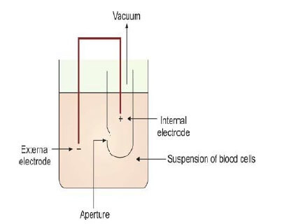
The electrical impedance method, often referred to as the Coulter counter method, is a well-established technique used for counting the cellular components of blood. This principle, initially developed by Coulter Electronics, relies on the interruption of an electrical current as cells pass through a small aperture between two electrodes. These electrodes are immersed in an isotonic solution, and as a cell moves through the aperture, it impedes the current, creating a voltage pulse. This pulse is proportional to the size of the cell and is measured to determine its volume.

For accurate cell counting, the blood sample must be diluted so that only a few cells pass through the aperture at once. The electrical impedance method works by amplifying the pulse generated when a cell crosses the aperture. The duration of the pulse reflects the time taken for the cell to pass through, while its height is directly related to the cell's size. Some hematology analyzers employ hydrodynamic focusing to ensure that each cell follows the same central path, reducing inaccuracies in volume measurement caused by cells moving off-center. This process is key in CBC test machines for determining blood cell counts and platelet counts.
In a typical setup, a blood sample treated with an anticoagulant is drawn into the analyzer and divided into two parts. One portion is directed to measure red blood cells and platelets, while the other focuses on white blood cells (WBCs) after lysing the red cells. Platelets are counted based on particles between 2-20 femtoliters, while red cells are within the 36-360 femtoliter range. Hemoglobin concentration is estimated through light transmission at 535 nm, a vital aspect of blood analysis.
2. Light Scatter
In automated hematology analyzers, light scatter is utilized to differentiate cell types based on their physical characteristics. Cells flow individually through a flow cell, and a laser beam targets them. As the light hits each cell, it scatters in different directions. The forward scatter (FALS) corresponds to the cell's size, while the side scatter (SS) at a 90° angle gives information about the cell's internal complexity, such as nuclear density and cytoplasmic granularity. This dual measurement allows for the clear distinction between granulocytes, lymphocytes, and monocytes.
3. Fluorescence
Fluorescence technology in hematology analyzers measures the cellular content of RNA, DNA, and cell surface antigens. For example, reticulocytes (immature red blood cells) are identified by their RNA content, while nucleated red cells are detected through their DNA fluorescence. This method provides an additional layer of specificity, especially in identifying certain subpopulations of blood cells.
4. Light Absorption
The concentration of hemoglobin is typically measured through light absorption using spectrophotometry. Hemoglobin is converted into cyanmethemoglobin, or a similar compound, before absorbance is measured. Some hematology analyzers also classify white blood cells based on peroxidase activity, which is detected through absorbance. This method is vital for accurate hemoglobin estimation and leukocyte classification.
5. Electrical Conductivity
In addition to electrical impedance, certain analyzers utilize electrical conductivity to classify leukocytes. By passing a high-frequency current through the cells, the analyzer can determine their physical and chemical properties, which aids in more precise classification of white blood cells.
These principles form the foundation of modern hematology analyzers and contribute to their widespread use in medical laboratories worldwide. They enable rapid, accurate cell counts and measurements, essential for diagnostic testing such as complete blood counts (CBCs).
The information on this page is peer reviewed by a qualified editorial review board member. Learn more about us and our editorial process.
Last reviewed on .
Article history
- Latest version
Cite this page:
- Posted by Dayyal Dungrela