Hidden Blood-Making Organ Discovered at the Maternal-Fetal Boundary
Researchers identify a previously unknown extraembryonic tissue that generates specialized immune and blood cells long before stem cells emerge.
Hidden blood-making machinery tucked inside the maternal-fetal boundary in mice and humans has just been uncovered, rewriting foundational textbook models of embryonic development. For decades, scientists believed that all early blood production before the arrival of stem cells occurred exclusively inside the extraembryonic yolk sac. Now, a multidisciplinary research team has identified a previously unknown hematopoietic site at the implantation zone, challenging long-held assumptions about where our earliest immune cells and red blood cells originate.
What is the MIXER Tissue and Where is it Located?
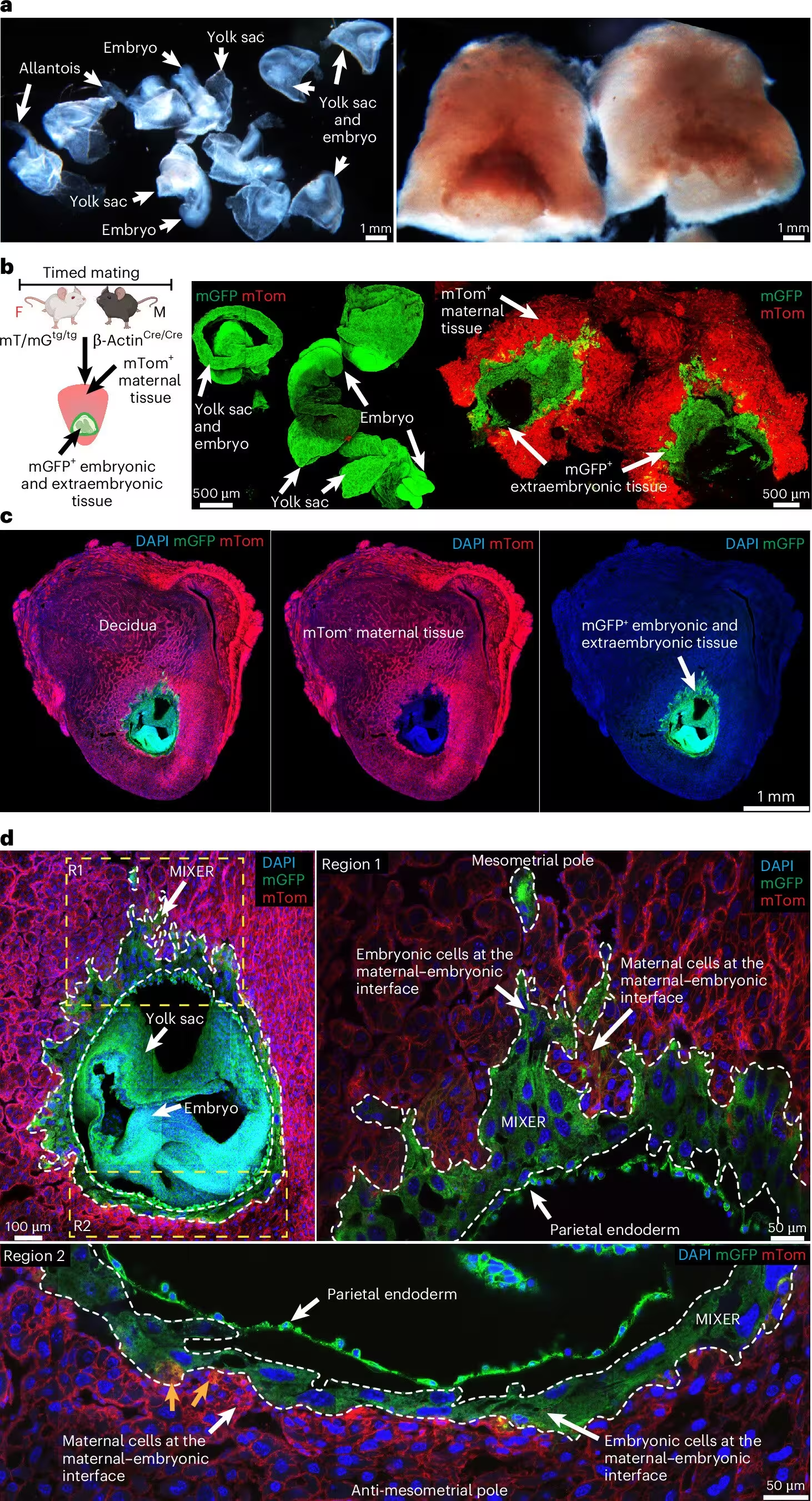
The newly discovered compartment has been named MIXER, which stands for Maternally Interfaced eXtraEmbRyonic tissue. Situated right at the site of embryo implantation in the uterus, MIXER develops at the exact same time as the yolk sac during early pregnancy. Using sophisticated genetic mouse models that distinguish between maternal and embryonic cells through fluorescent proteins, the researchers found that embryonic cells actively invade the uterine wall at the mesometrial pole, forming a distinct vascularized bed that operates independently of the yolk sac. This study was published in Nature Immunology.
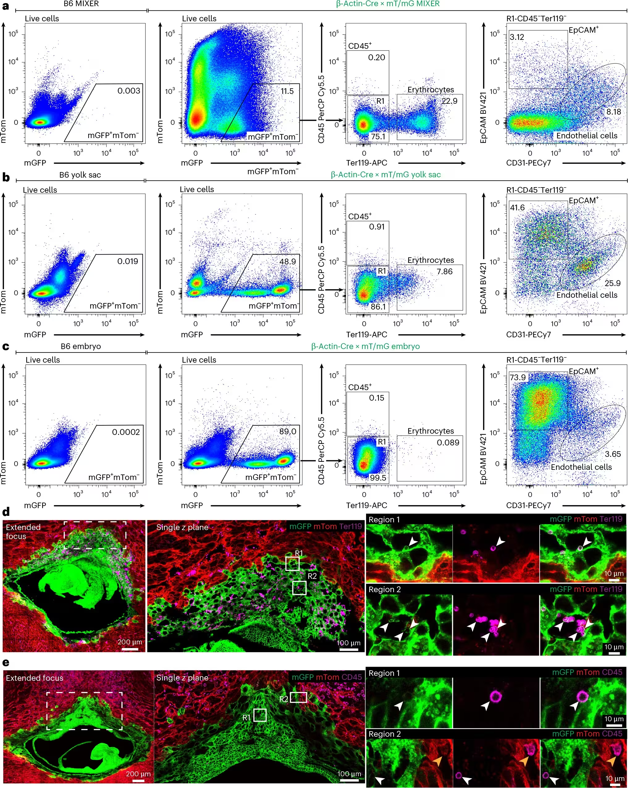
Advanced confocal imaging and flow cytometric screening revealed that MIXER is not merely a passive reservoir for blood cells spilling over from nearby tissues. Instead, it teems with embryonic endothelial cells, early erythrocytes, and leukocytes as early as embryonic day 7.5 and 8.5. The team detected robust vascular markers such as CD31 and CD309 within the tissue, confirming that a local microenvironment exists to support blood development well before the embryo establishes a full circulatory loop with its extraembryonic membranes.
Which Unique Progenitor Cells Reside Inside MIXER?
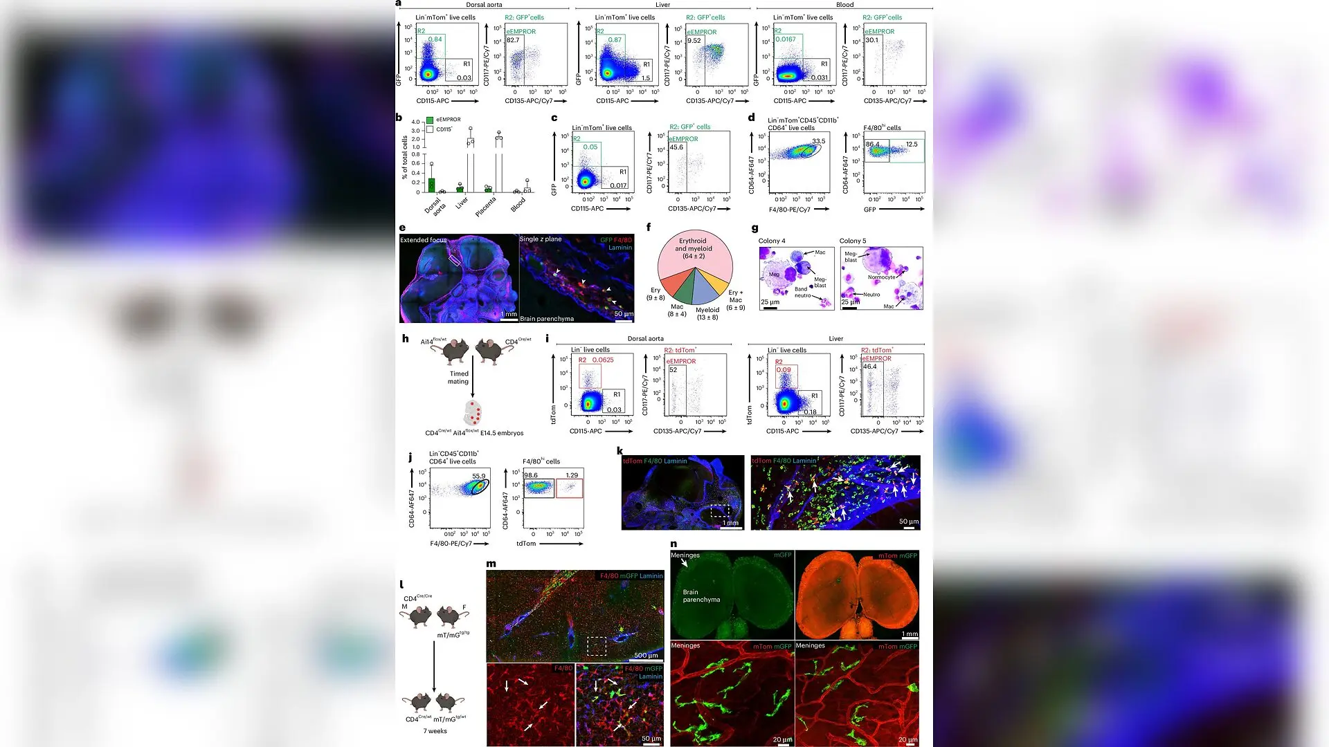
While the yolk sac is known for producing classical erythro-myeloid progenitors, which are specialized precursor cells that generate long-lived tissue-resident macrophages like microglia in the brain and Kupffer cells in the liver, MIXER contains something entirely unexpected. In addition to abundant classical colony-stimulating factor 1 receptor-positive progenitors, MIXER harbors a distinct population of CD4-expressing embryonic erythro-myeloid progenitors dubbed eEMPROR cells. These cells are completely absent from the yolk sac.
When isolated and cocultured with supportive stromal cells or grown in specialized media, these MIXER-derived progenitors demonstrated a powerful capacity to differentiate into multiple blood lineages, including erythroid, myeloid, and megakaryocytic cells. Lineage-tracing experiments in mice confirmed that these CD4-marked progenitors do not simply vanish after early gestation. They migrate into the developing embryo and give rise to specialized macrophage populations, contributing heavily to the perivascular space around the brain, meninges, skin, and liver, where some descendants persist all the way into adulthood.
Does This Maternal-Fetal Blood Network Also Exist in Humans?
To determine whether these findings extend beyond rodents, the investigators analyzed first-trimester human placental and decidual tissues collected at approximately five weeks of gestation. Immunofluorescence staining of these early human samples revealed discrete cellular clusters of CD4-positive cells encapsulated within CD31-positive endothelial-lined structures inside the villous core. Crucially, these CD4-positive cells did not express leukocyte markers like CD45 or macrophage markers like CD14, closely mirroring the molecular profile of the eEMPROR lineage found in mice. Furthermore, the presence of nucleated CD235a-positive erythroid cells confirmed that active blood formation is underway in early human placental villi long before fetal hematopoietic stem cells emerge.
The discovery of MIXER helps explain persistent blind spots in developmental biology, where implantation-site tissues were frequently discarded or overlooked due to technical difficulties in separating maternal and embryonic compartments. By revealing a dual-territory origin for early blood development, the work opens new avenues for exploring how disruptions at the maternal-fetal interface might influence lifelong immune health, neurological development, and disease susceptibility.
The research was published in Nature Immunology on November 1, 2026.
This article has been fact checked for accuracy, with information verified against reputable sources. Learn more about us and our editorial process.
Last reviewed on .
Article history
- Latest version
Reference(s)
- Jain, Rohit., et al. “The maternal–fetal interface is an early hematopoietic niche enriched in distinct erythro-myeloid progenitors.” Nature Immunology, vol. 27, no. 11, October 26, 2026, pp. 2306-2321. Springer Science and Business Media LLC, doi: 10.1038/s41590-026-02683-4. <https://doi.org/10.1038/s41590-026-02683-4>.
Cite this page:
- Posted by Faisal Qureshi