Bacteria Use a Helical Moving Walkway to Glide Across Surfaces
High-resolution imaging reveals how inner-membrane motors power a dynamic protein belt to drive bacterial movement.

Bacteria rely on a dynamic moving walkway beneath their outer membrane to glide across solid surfaces without the aid of flagella or pili. In a study published in Science Advances, researchers isolated and imaged the helical filament that drives this movement in the gliding bacterium Flavobacterium johnsoniae. The findings reveal how cellular machinery couples protein secretion directly with rapid surface translation.
Unlocking the Structure of the Gliding Belt at High Resolution
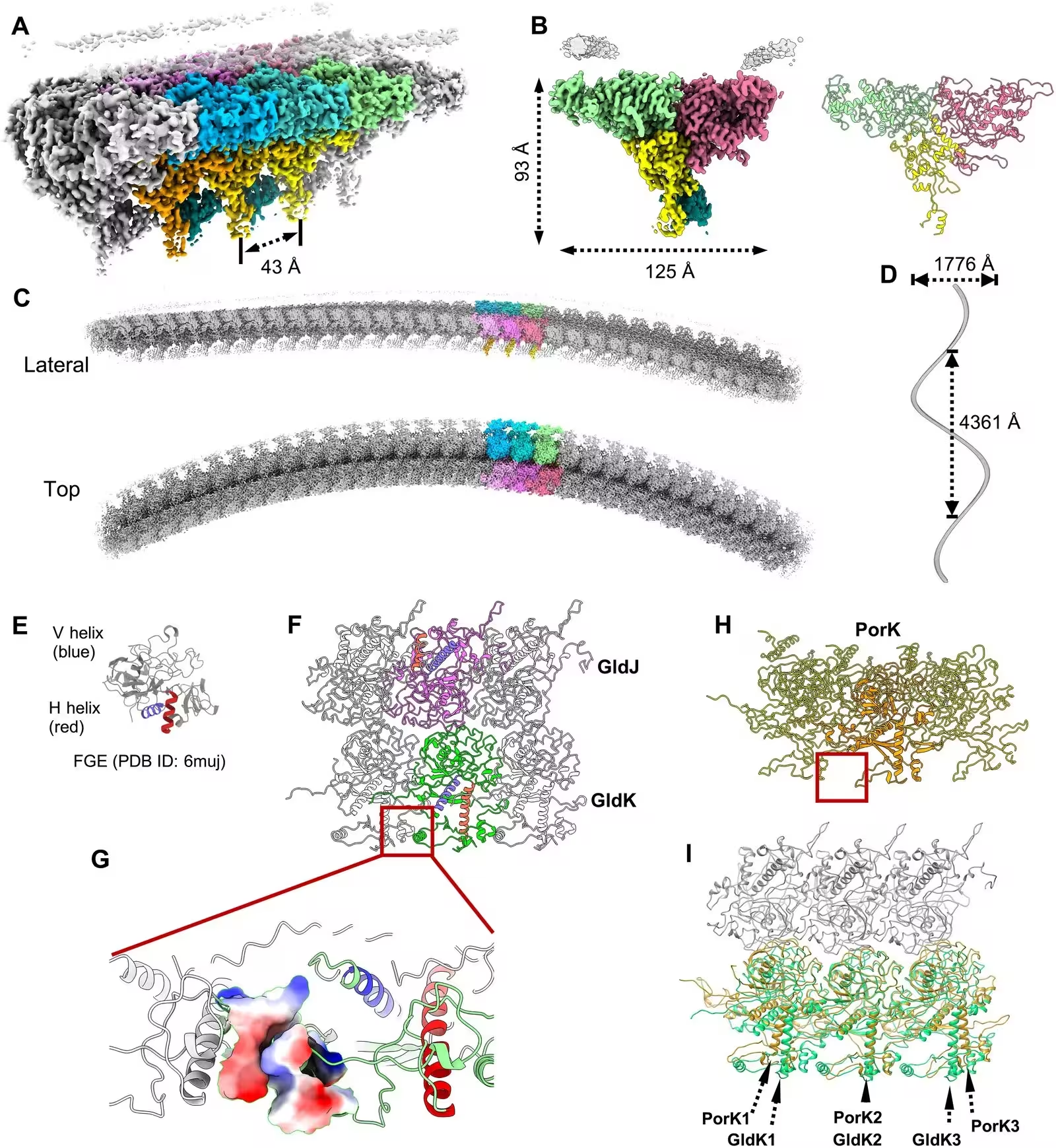
Many rod-shaped bacteria belonging to the phylum Bacteroidota move across surfaces by secreting surface adhesins that travel in helical paths along the cell body. Researchers used a streamlined purification strategy involving detergent solubilization and differential centrifugation to isolate these elusive filaments. Analyzing the purified sample using single-particle cryo-electron microscopy, the team reconstructed the filament backbone at a resolution of 2.63 angstroms.
The structural analysis showed that the filament consists of repeating trimeric units containing one copy each of the core subunits GldJ, GldK, and GldN. Together, these proteins form an asymmetric prism-shaped block measuring approximately 125 angstroms wide and 93 angstroms high. The lipoproteins GldJ and GldK sit side by side beneath the outer membrane, while GldN occupies the central cavity and extends into the periplasm.
How Molecular Motors Power the Moving Walkway
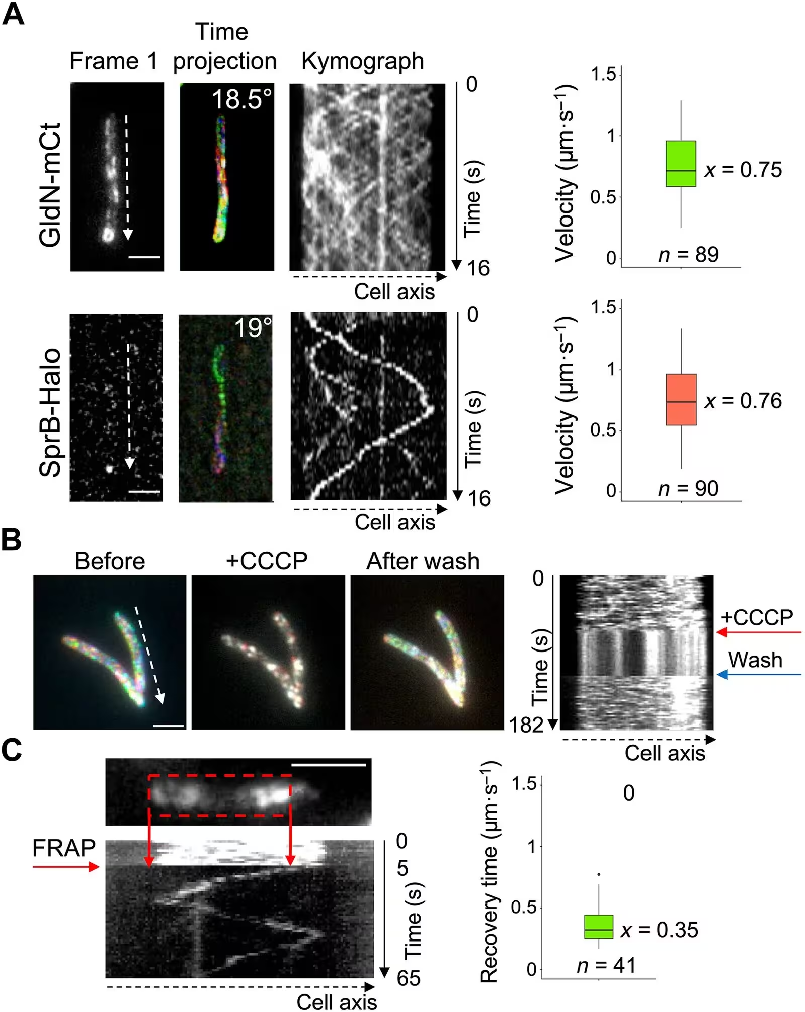
Further investigation demonstrated that the GldJKN units assemble into a right-handed helix with a twist of roughly 2.4 degrees per unit. Live-cell imaging using fluorescent tags revealed that these helical belts are highly dynamic, traveling continuously from one cell pole to the other at speeds of approximately 0.75 micrometers per second. The major cell-surface adhesin, known as SprB, moves along the cell at a nearly identical speed and tilt angle.
The researchers observed that this continuous motion depends entirely on proton motive force dependent GldLM molecular motors embedded in the inner membrane. When the team injected a drug called CCCP to dissipate the proton gradient, the movement of the fluorescently labeled GldN-mCt patches halted immediately. Once the drug was washed out, the dynamic gliding motion rapidly resumed, confirming that motor activity provides the necessary mechanical torque to drive the belt.
Connecting Secretion to Surface Movement
The study provides a structural explanation for how bacteria coordinate protein export with whole-cell locomotion. The type IX secretion system typically transports proteins across the bacterial envelope, but in gliding species, this machinery appears repurposed. The shuttle protein SprF, which binds the adhesin SprB, forms a covalent disulfide bridge with the GldK lipoprotein, physically linking cargo delivery to the moving belt.
While the data point strongly to a coordinated moving walkway, open questions remain regarding whether the belt forms a single continuous structure or consists of shorter, interconnected segments that work together. Understanding the physical properties of the outer membrane and its specialized lipid composition will be essential for explaining how such rapid protein movement is sustained without disrupting the bacterial envelope.
The research was published in Science advances on November 18, 2026.
This article has been fact checked for accuracy, with information verified against reputable sources. Learn more about us and our editorial process.
Last reviewed on .
Article history
- Latest version
Reference(s)
- Liu, Hanxiong., et al. “Structure and dynamics of the cellular belt driving bacterial gliding motility.” Science Advances, vol. 12, no. 47, November 20, 2026 American Association for the Advancement of Science (AAAS), doi: 10.1126/sciadv.aef5514. <https://doi.org/10.1126/sciadv.aef5514>.
Cite this page:
- Posted by Elizabeth Taylor