Housekeeping Protein Drives Lung Cancer Identity
Unbiased gene-disruption screens reveal that a ubiquitous cellular factor teams up with master regulators to control tumor antigen production.
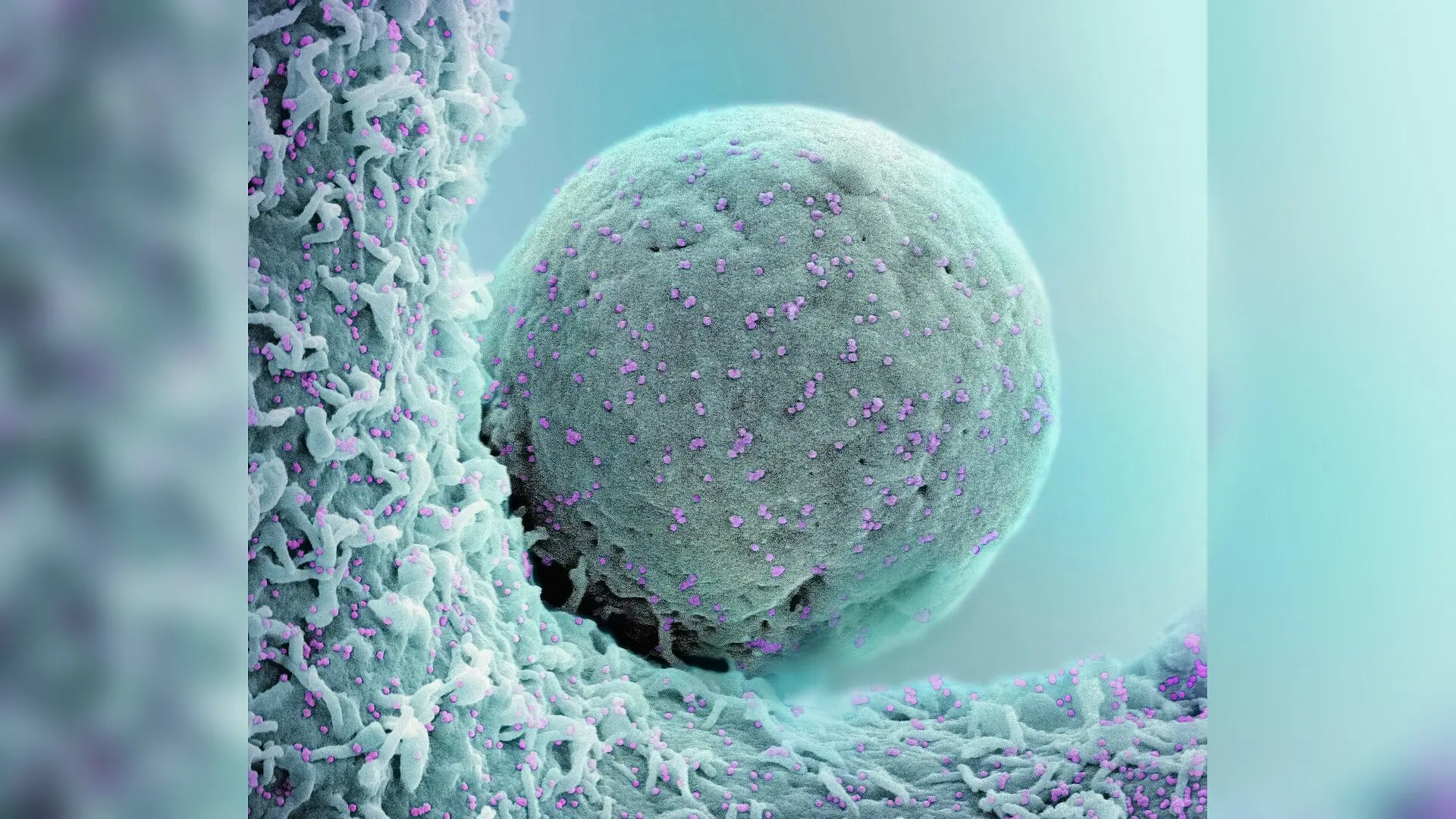
A widely distributed human protein usually tasked with basic cellular housekeeping has been unmasked as an unexpected driving force behind small cell lung cancer. Researchers mapping genetic dependencies in aggressive pulmonary tumors discovered that the transcription factor POU2F1 works in tandem with specialized master regulators to switch on genes that define the disease’s malignant identity.
Small cell lung cancer is a fast-growing form of cancer that forms distinct neuroendocrine characteristics, meaning the malignant cells adopt traits typical of hormone-producing nerve tissues. A hallmark of this state is the heavy production of a surface protein called delta-like ligand 3, or DLL3. Because this antigen appears almost exclusively on these tumor cells while remaining mostly absent in healthy adult organs, it serves as a primary target for specialized immunotherapies and antibody treatments.
Despite the critical importance of DLL3 in diagnosing and treating the disease, scientists have long debated the exact genetic mechanisms that prompt cancer cells to manufacture it in such large amounts. Previous assumptions tied the protein’s presence loosely to broader tumor subtypes, leaving the precise regulatory machinery hidden.
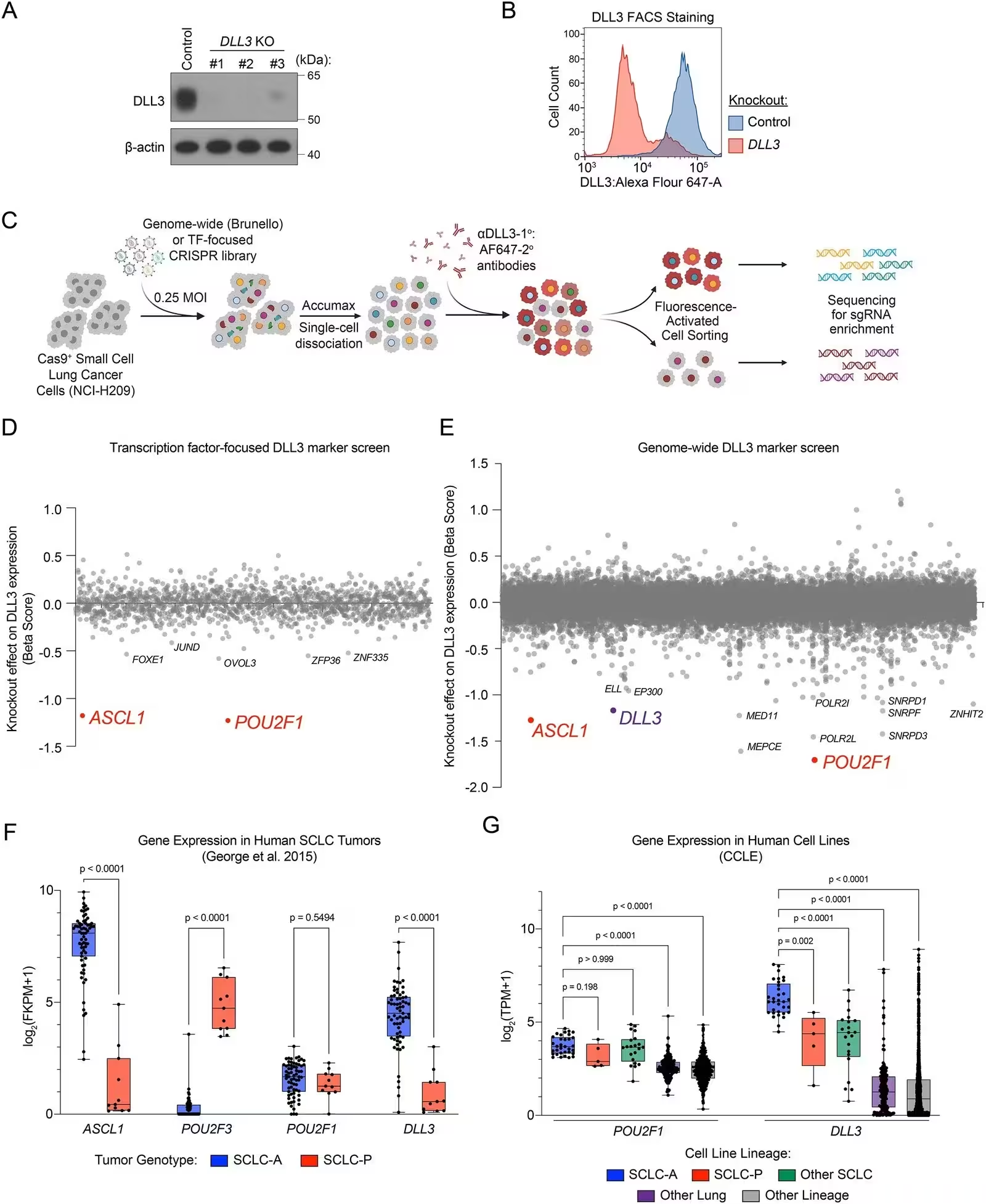
To uncover the drivers behind this process, a research team led by scientists at Cold Spring Harbor Laboratory conducted systematic genetic screening experiments using molecular disruption tools. Deploying both targeted transcription factor assays and genome-wide CRISPR screens in human cancer cell lines, the investigators systematically knocked out thousands of genes to see which ones caused DLL3 production to drop. The screening data, published in NAR Cancer, pointed directly to POU2F1 as a top activator.
How a General Housekeeping Factor Acquires Specialized Tumor Roles
POU2F1 is a ubiquitously expressed protein found across nearly all tissues in the human body, where it typically manages basal promoter activity, cellular metabolism, and stress responses. Because it is active everywhere, its identification as a specialized driver of lung cancer traits came as a surprise. Further investigation revealed that while the protein is present in many cells, it reaches unusually high expression levels in small cell lung cancer compared to other tumor types.
To understand how a general protein achieves such cancer-specific control, the team performed chromatin immunoprecipitation sequencing to map where the protein binds across the genome. The mapping data showed that POU2F1 does not work alone. Instead, it converges on specific genetic regulatory regions alongside ASCL1, a well-known master regulator that drives neuroendocrine identity in these tumors.
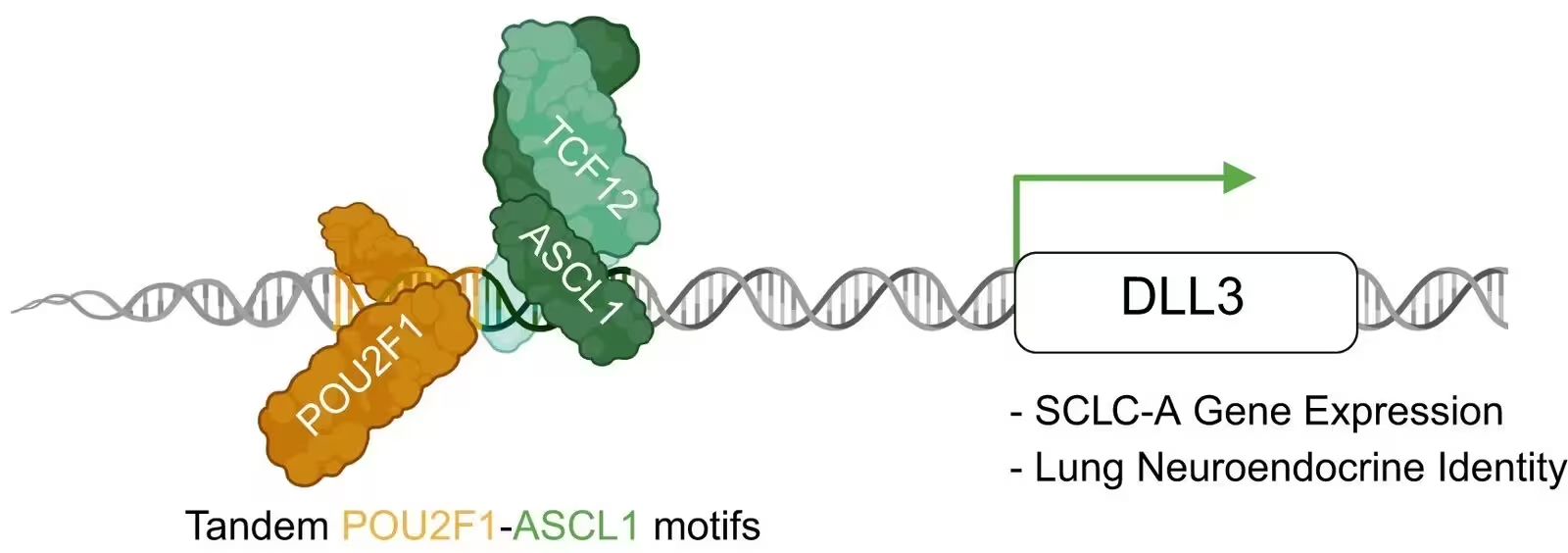
Detailed sequence analysis of the DLL3 promoter revealed a specific arrangement featuring adjacent binding sites for both proteins, known as tandem POU2F1 and ASCL1 motifs. When the researchers mutated these motifs or reversed their orientation in laboratory assays, the cooperative activation of the DLL3 promoter was completely abolished, proving that the precise spatial layout of the DNA sequence is mandatory for the partnership to function.
Structural Models Reveal Molecular Contact Points
To visualize how these proteins physically interact on the DNA strand, the investigators utilized advanced computational structural prediction models via AlphaFold 3. The generated simulations indicated that the DNA double helix acts as a stabilizing scaffold, facilitating direct physical contact between POU2F1 and an accompanying protein complex containing ASCL1 and TCF12.
When the computer models simulated a reversed DNA motif or removed the DNA strand entirely, the structural confidence and stability of the protein interactions dropped markedly. This finding supports a hierarchical model of gene activation where the master regulator ASCL1 first engages closed chromatin to open up access, allowing POU2F1 to dock securely into the composite motif and drive robust transcription.
Broader Implications for Tumor Plasticity and Therapy
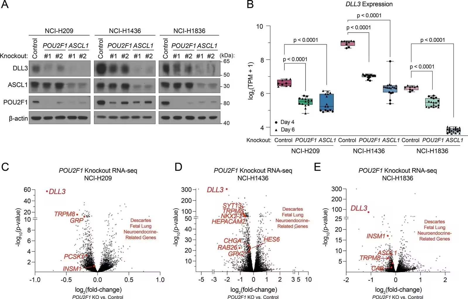
Expanding their genome-wide analysis beyond just a single gene, the team scanned the entire human genome for similar tandem sequence motifs. They identified hundreds of comparable composite elements located near genes tied specifically to lung neuroendocrine cell functions. Subsequent RNA sequencing after knocking out POU2F1 confirmed that a wide network of neuroendocrine identity markers experienced significant downregulation.
The authors note that these insights provide a foundation for understanding tumor plasticity, or the ability of cancer cells to shift between different molecular states to evade treatment. While pharmacological manipulation of this protein interaction remains speculative at this stage, understanding the foundational grammar of tumor-associated antigens offers new perspectives for stabilizing targeted therapies and preventing resistance.
The research was published in NAR cancer on November 18, 2026.
This article has been fact checked for accuracy, with information verified against reputable sources. Learn more about us and our editorial process.
Last reviewed on .
Article history
- Latest version
Reference(s)
- Cunniff, Patrick J., et al. “Marker-based CRISPR screens identify POU2F1 as a regulator of DLL3 and neuroendocrine identity in small cell lung cancer.” NAR Cancer, vol. 8, no. 4, November 18, 2026 Oxford University Press (OUP), doi: 10.1093/narcan/zcag032. <https://doi.org/10.1093/narcan/zcag032>.
Cite this page:
- Posted by Faisal Qureshi