Scientists Find Potential Antiviral Defense Against Deadly Tick Virus
High-throughput screening of nearly 109,000 compounds reveals small molecules that shut down viral protein machinery in laboratory models.
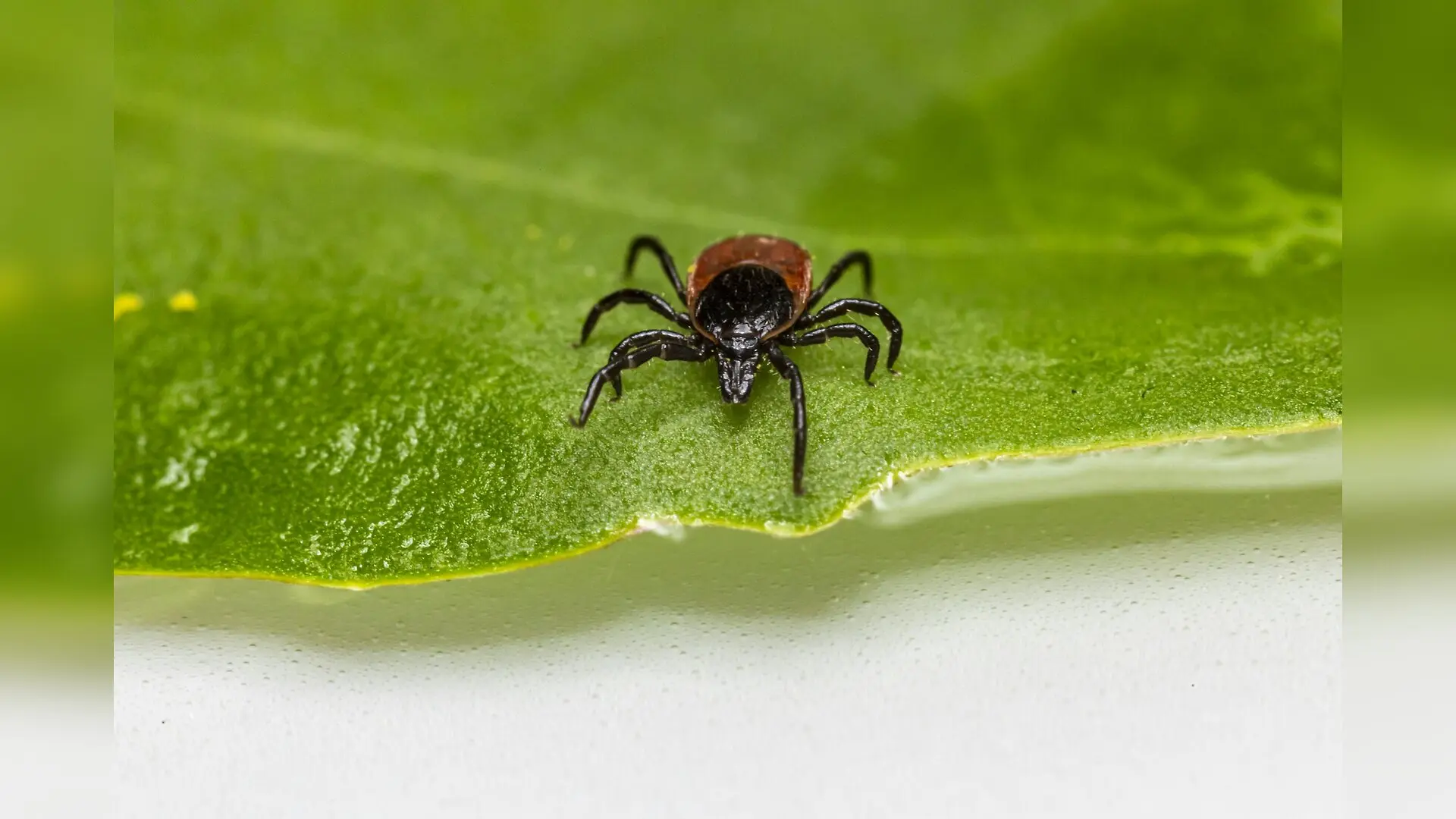
Tiny tick-borne pathogens continue to threaten global health, and researchers have just uncovered a chemical vulnerability in the deadly Crimean-Congo hemorrhagic fever virus. Scientists identified three small molecules that block a crucial viral protein from hijacking human cell machinery, potentially opening the door to the first targeted antiviral treatment for the disease.
How a Deadly Virus Hijacks Human Cells
Crimean-Congo hemorrhagic fever is a severe viral illness transmitted primarily through the bites of infected ticks or contact with infected livestock blood. The disease causes sudden high fever, muscle aches, and dangerous internal bleeding, with a case fatality rate ranging between 10% and 40%. Because no approved vaccines or specific antiviral treatments currently exist, doctors rely entirely on supportive care. Global climate shifts and expanding livestock trade have pushed tick populations into new regions, raising urgent concerns about the spread of the pathogen.
To replicate inside a host, the virus relies on its nucleocapsid protein, a multifunctional building block that packages the viral genetic material and helps manufacture viral proteins. Researchers recently published a detailed description of these screening methods and molecular interactions published in PLOS Pathogens. The study shows that the viral nucleocapsid protein binds to a specific hairpin structure at the front end of the viral messenger RNA. By anchoring itself simultaneously to this viral RNA structure and to a human cellular translation factor known as eIF4A, the viral protein forces the host cell to manufacture viral proteins in large quantities.
Screening Over 100,000 Chemical Compounds
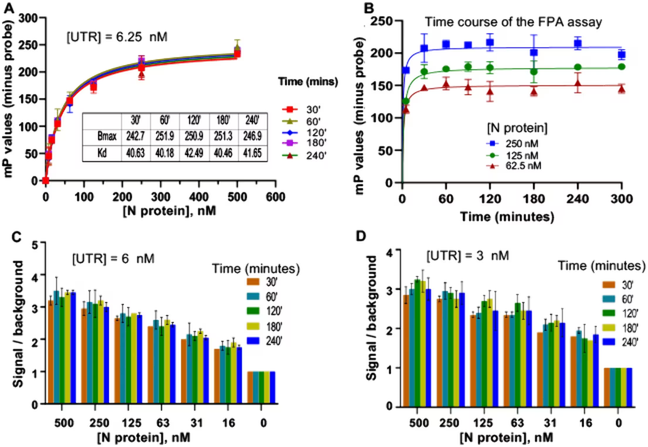
To interrupt this protein-RNA partnership, the research team developed a high-throughput fluorescence-based assay capable of measuring molecular binding with high precision. This technology enabled the team to screen a massive chemical library containing 108,947 distinct small molecules. The primary screen flagged 122 candidate compounds that interfered with the protein-RNA interaction. Subsequent dose-response evaluations and chemical clustering narrowed the selection down to a group of high-confidence candidates, focusing particularly on three compounds designated as compound 11, compound 14, and compound 33.
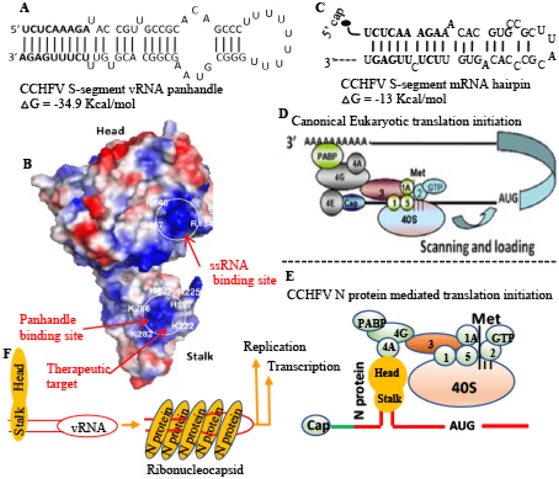
Biochemical assays demonstrated that these three agents disrupted the binding between the viral protein and the messenger RNA hairpin with half-maximal inhibitory concentration values ranging from 11 to 41 micromolars. Further mechanistic tests revealed that these compounds act as noncompetitive inhibitors. Instead of blocking the RNA-binding groove directly, they bind to a distinct pocket on the stalk domain of the viral protein, inducing conformational changes that prevent proper engagement with the viral RNA.
Testing Antiviral Efficacy in Laboratory Models
Because working with the live Crimean-Congo hemorrhagic fever pathogen requires maximum-security containment facilities, the team tested the antiviral efficacy of the compounds using the Hazara virus, a related lesser-known virus that serves as a safe and reliable laboratory surrogate. Structural comparisons of the stalk domains between the two viruses confirmed a high degree of architectural conservation, validating the surrogate model.
When infected mammalian cells were treated with the candidate compounds, viral replication dropped significantly. Compounds 11 and 14 reduced the release of infectious viral particles into the culture medium by more than 100-fold, while compound 33 suppressed viral progeny to nearly undetectable levels. Cytotoxicity tests across multiple human and animal cell lines showed that these molecules caused very little cellular damage at effective therapeutic doses, yielding high selectivity indices that favor safety.
Mutations Reveal Where the Inhibitors Bind
To pinpoint exactly how compound 11 exerts its effect, the researchers engineered specific mutations into the viral protein’s stalk domain, substituting key amino acids such as lysine, glutamine, and glutamic acid with alanine. While these mutated proteins retained their normal ability to bind viral RNA, compound 11 completely lost its inhibitory effect against the modified proteins. This genetic mapping confirmed that specific residues including Lys473, Gln300, and Glu240 form the essential binding site for compound 11. In contrast, compounds 14 and 33 retained their inhibitory activity against the mutants, suggesting they operate through slightly different interacting sites elsewhere on the protein.
While these laboratory findings offer a promising foundation for drug development, several hurdles remain before human therapies can be tested. Researchers must still confirm whether the inhibitor-binding pockets remain exposed and vulnerable within fully assembled viral ribonucleoprotein complexes inside infected hosts. Future investigations will focus on chemical optimization of these molecular scaffolds, co-crystallization studies to map atomic interactions, and pharmacokinetic testing in animal models.
The research was published in PLOS Pathogens on November 20, 2026.
This article has been fact checked for accuracy, with information verified against reputable sources. Learn more about us and our editorial process.
Last reviewed on .
Article history
- Latest version
Reference(s)
- Le, Hoangvi., et al. “A new therapeutic opportunity in Crimean-Congo hemorrhagic fever: Targeting the stalk domain of viral nucleocapsid protein for therapeutic intervention.” PLOS Pathogens, vol. 22, no. 11, November 20, 2026, pp. e1014772 Public Library of Science (PLoS), doi: 10.1371/journal.ppat.1014772. <https://doi.org/10.1371/journal.ppat.1014772>.
- Posted by Elizabeth Taylor