Novel Glycans Shield Insulin-Secreting Cells From Destructive Diabetes Inflammation
Modified aminoglycoside compounds neutralize a key enzyme that strips away protective sugar coatings and triggers cell death in human pancreatic islets.
Synthetic molecules derived from an old antibiotic can protect delicate insulin-producing cells from destructive inflammation and oxidative damage, offering fresh hope for diabetes intervention. The research, published in ACS Pharmacology & Translational Science, demonstrates that targeting a specific enzyme involved in tissue degradation can interrupt the cascading cellular injury characteristic of early diabetes progression.
How an Enzyme Drives Inflammation and Cell Damage
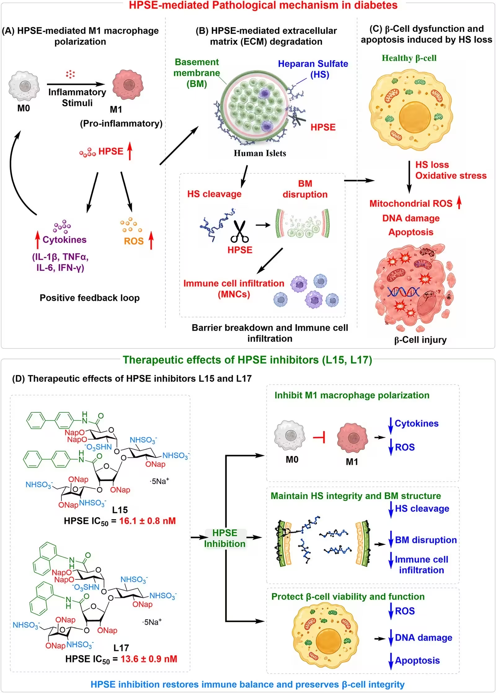
During the onset of type 1 diabetes, pancreatic beta-cells face a relentless onslaught from the immune system. Beyond direct autoimmune attacks, specialized immune cells called macrophages infiltrate the pancreas and release an enzyme known as heparanase, or HPSE. This enzyme normally chews up heparan sulfate, a protective sugar coating that maintains the structural integrity of the extracellular matrix surrounding insulin-producing cell clusters known as islets. When heparanase strips away this protective coating, it triggers a destructive cycle of inflammation, oxidative stress, and eventual cell death.
To combat this pathway, a team of researchers led by scientists at Wayne State University developed two specialized drug candidates named L15 and L17. These compounds act as artificial mimetics of the body’s natural sugar chains, binding directly to heparanase and blocking its destructive enzymatic activity. Unlike older experimental drugs that required dozens of complicated chemical steps to manufacture, these new aminoglycoside derivatives can be synthesized far more efficiently, making them practical candidates for therapeutic development.
What Happens When Inhibitors Block the Damage Cascade
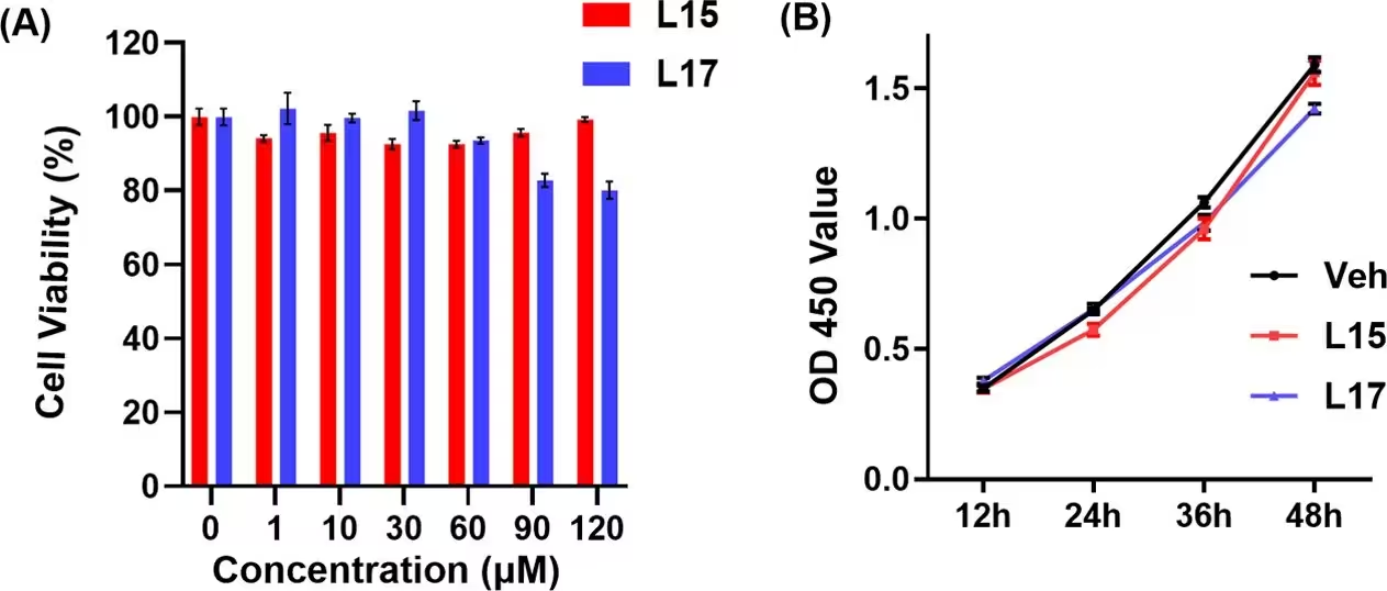
In laboratory experiments using mouse macrophages, pretreating the cells with L15 or L17 significantly suppressed the activation of pro-inflammatory immune pathways. The treatments dramatically reduced the expression of inflammatory markers like inducible nitric oxide synthase and key signaling proteins including tumor necrosis factor alpha and interleukin-6. When these treated immune secretions were applied to pancreatic beta-cell models, the protective compounds successfully preserved cell viability and prevented the structural collapse typically caused by inflammatory stress.
Further microscopic analysis revealed the precise timeline of cellular destruction. The team discovered that mitochondrial oxidative stress, marked by a surge in reactive oxygen species, begins as early as 8 hours after exposure to inflammatory triggers. This oxidative stress acts as an upstream driver that subsequently ruins the cellular cytoskeleton by dismantling F-actin networks before finally inducing programmed cell death 24 hours later. By introducing the antioxidant N-acetylcysteine or applying the heparanase inhibitors L15 and L17, the researchers successfully halted this destructive chain reaction.
Protecting Human Islets Ex Vivo
To verify whether these laboratory findings translate to human biology, the investigators tested the compounds on human pancreatic islets obtained from cadaveric donors. When untreated human islets were exposed to heparanase and inflammatory cytokines, they suffered a near-complete loss of their protective heparan sulfate coating, experienced massive oxidative stress, and lost their ability to secrete insulin in response to changing glucose levels.
Can L17 Restore Normal Insulin Secretion?
When human islets were treated with compound L15 or L17 alongside inflammatory triggers, the results were strikingly positive. Both molecules preserved the delicate heparan sulfate coating and suppressed total reactive oxygen species. Compound L17 proved particularly potent, consistently restoring glucose-stimulated insulin secretion to levels closely matching healthy, untreated control islets. These findings suggest that blocking heparanase shields human tissue from inflammatory degradation and maintains vital metabolic functions under stress.
While these initial ex vivo results mark an encouraging step forward, the authors note that further validation in living animal models of diabetes, such as mice treated with streptozotocin or non-obese diabetic strains, will be essential. Future studies will determine whether these protective mechanisms translate into sustained glycemic control and delayed disease progression in whole organisms.
The research was published in ACS pharmacology & translational science on October 5, 2026.
This article has been fact checked for accuracy, with information verified against reputable sources. Learn more about us and our editorial process.
Last reviewed on .
Article history
- Latest version
Reference(s)
- Wang, Junzhe., et al. “Heparanase-Inhibiting Glycans Protect Pancreatic β-Cells from Oxidative and Inflammatory Injuries and Preserve Insulin Production in Human Islets.” ACS Pharmacology & Translational Science, vol. 9, no. 11, October 5, 2026, pp. 3203-3216. American Chemical Society (ACS), doi: 10.1021/acsptsci.6c00370. <https://doi.org/10.1021/acsptsci.6c00370>.
Cite this page:
- Posted by Faisal Qureshi