New AI Framework Connects Six Layers of Cancer Data
By organizing molecular data into biological hierarchies and reconstructing missing records, a new computational model maps patient survival risks and core tumor drivers.

A new artificial intelligence framework successfully integrates six distinct types of biological data to map cancer vulnerabilities while reconstructing missing patient information. The computational system, developed by researchers and available on the arXiv preprint server, tackles one of modern oncology’s biggest hurdles by finding hidden patterns across massive amounts of medical data.
Precision medicine relies on combining massive amounts of biological information, from genetic mutations down to microscopic tissue images. However, most computer models treat these different data layers as flat files or lose vital connections between them, making it difficult to understand how a tumor actually grows. Furthermore, real-world hospital databases frequently suffer from incomplete records, where doctors have some test results for a patient but are missing others.
To overcome these hurdles, the research team designed an advanced computational architecture named Hierarchical Interpretable Tensor Fusion, or HIT-Fuse. Instead of dumping all patient data into a single pile, the system groups information according to the central dogma of molecular biology. It separates data into a DNA layer covering somatic copy number variations and DNA methylation, an RNA layer capturing gene and microRNA expression, and a functional expression layer combining protein abundance with digital pathology images extracted from whole-slide tissue scans.
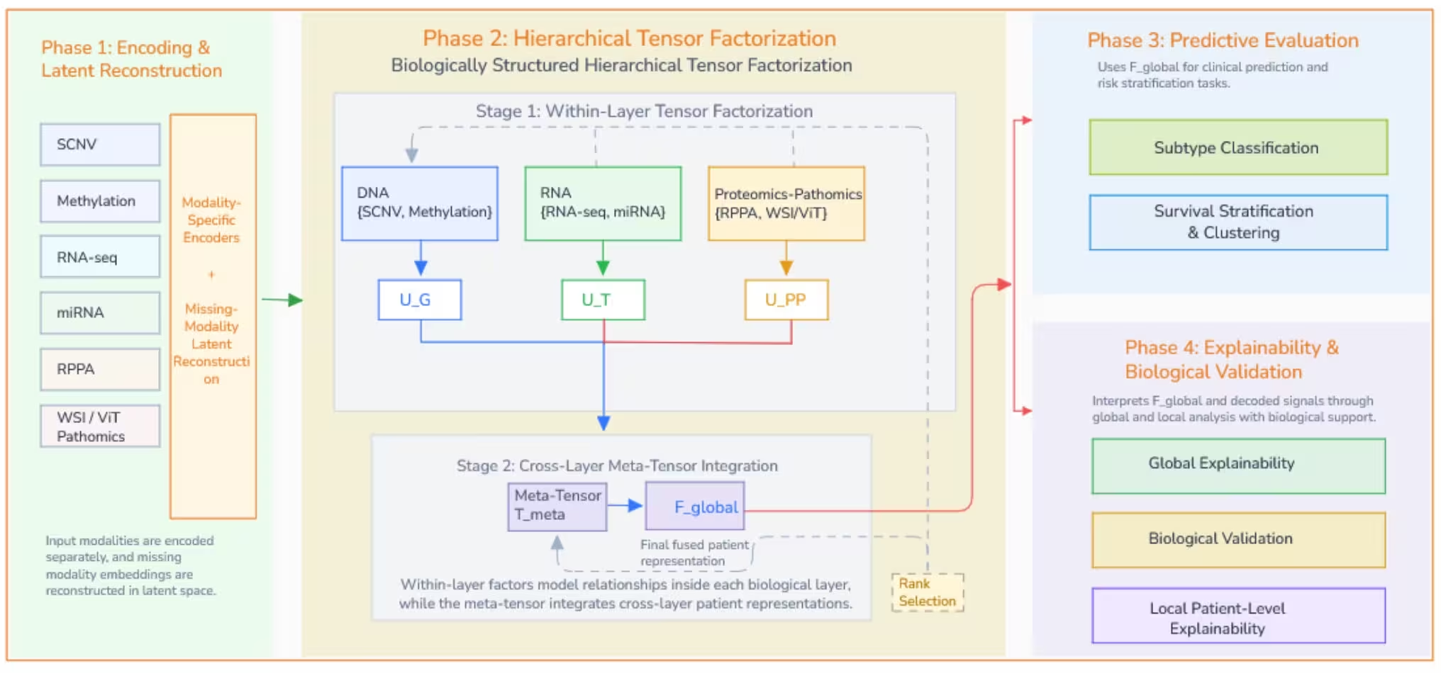
How Multi-Layer Tensor Decompositions Map Tumors
The system uses specialized mathematical tools known as tensor decompositions to model multi-way relationships between patients, data modalities, and biological factors. By organizing data into multi-dimensional arrays, the model can look at within-layer interactions before evaluating cross-layer connections. This hierarchical structure mirrors the natural flow of biological information from genes to proteins and tissue phenotypes.
To solve the problem of missing hospital records, the authors incorporated a latent-space super-imputation mechanism. When a patient’s file lacks a specific molecular test, the system uses clinical covariates and the remaining available omics data to reconstruct the missing latent representations. This process expanded the study cohorts significantly, such as increasing the glioma cohort from 397 complete patients to 1,110 individuals, without destabilizing the mathematical framework.
Validating Discovered Drivers Against Biological Screens
The investigators tested their framework across four major cancer types from The Cancer Genome Atlas, including glioma, lung adenocarcinoma, breast invasive carcinoma, and kidney cancer. When evaluating prognostic power, the model achieved strong survival stratification, yielding concordance index values up to 0.752 in glioma and 0.722 in kidney cancer. In addition, clustering the learned latent space revealed distinct risk groups with statistically significant survival differences across the cohorts.
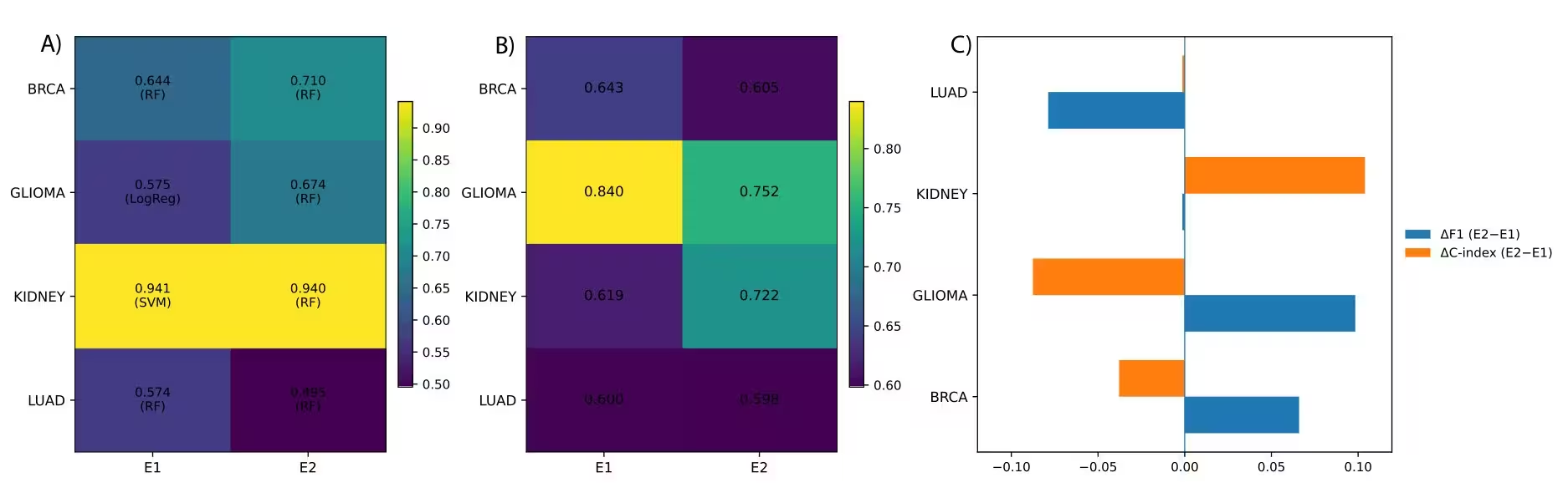
To confirm that the model was finding real biological signals rather than mathematical artifacts, the team traced high-level meta-factors back to raw features using a mathematical traceback mechanism. They compared these decoded molecular drivers against external functional databases, including CRISPR-Cas9 gene dependency screens from the Cancer Dependency Map and validated regulatory interactions from miRTarBase. The analysis showed that between 77 and 84 percent of selected genes matched DepMap matrices, and several identified drivers satisfied strict essentiality criteria in cancer-matched cell lines.
While simpler feature concatenation methods sometimes yielded higher classification accuracy scores in specific tests, the researchers note that flat models lack built-in interpretability. The hierarchical tensor design provides a transparent chain of attribution, allowing scientists to trace predictions from high-level clinical risk factors down to individual genes and tissue features.
The study was posted to the arXiv preprint server on November 19, 2026.
This article has been fact checked for accuracy, with information verified against reputable sources. Learn more about us and our editorial process.
Last reviewed on .
Article history
- Latest version
Reference(s)
- Hussein, Ahmad. “An Explainable and Scalable Hierarchical Tensor Framework for Integrating Multi-Omics and Digital Pathomics.” arXiv.org <http://arxiv.org/abs/2611.25584v1>.
Cite this page:
- Posted by Elizabeth Taylor