Targeted Light-Activated Nanoparticles Boost Chemotherapy Response in Pancreatic Cancer Models
Laboratory tests demonstrate that pairing light-triggered liposomes with chemotherapy yields strong synergistic inhibition and enhances immune signaling.

Targeted light-activated nanoparticles can overcome the stubborn resistance of pancreatic cancer by supercharging the immune-stimulating effects of standard chemotherapy, according to a laboratory study evaluating genetically engineered mouse models. Pancreatic ductal adenocarcinoma, a notoriously aggressive form of tumor, often fails to respond to standard combinations of chemotherapy and immunotherapy that succeed in other malignancies. To tackle this treatment resistance, researchers designed a dual-action system that combines light-triggered phototherapy with immune checkpoint blockade.
Led by Chanda Bhandari and Girgis Obaid from the University of Texas Southwestern Medical Center, the research team investigated whether pairing nanoparticle albumin-bound paclitaxel, a widely used chemotherapy drug commonly known as nab-PTX, with targeted photoactivatable liposomes could yield synergistic therapeutic benefits. The study is available on RSC Pharmaceutics, outlining a series of in vitro experiments across multiple mouse pancreatic cancer cell lines. While conventional treatments often struggle against the dense scar tissue characteristic of pancreatic tumors, this novel approach aims to prime the cellular environment and trigger robust immune responses.
How Do Light-Activated Nanoparticles Target Cancer Cells?
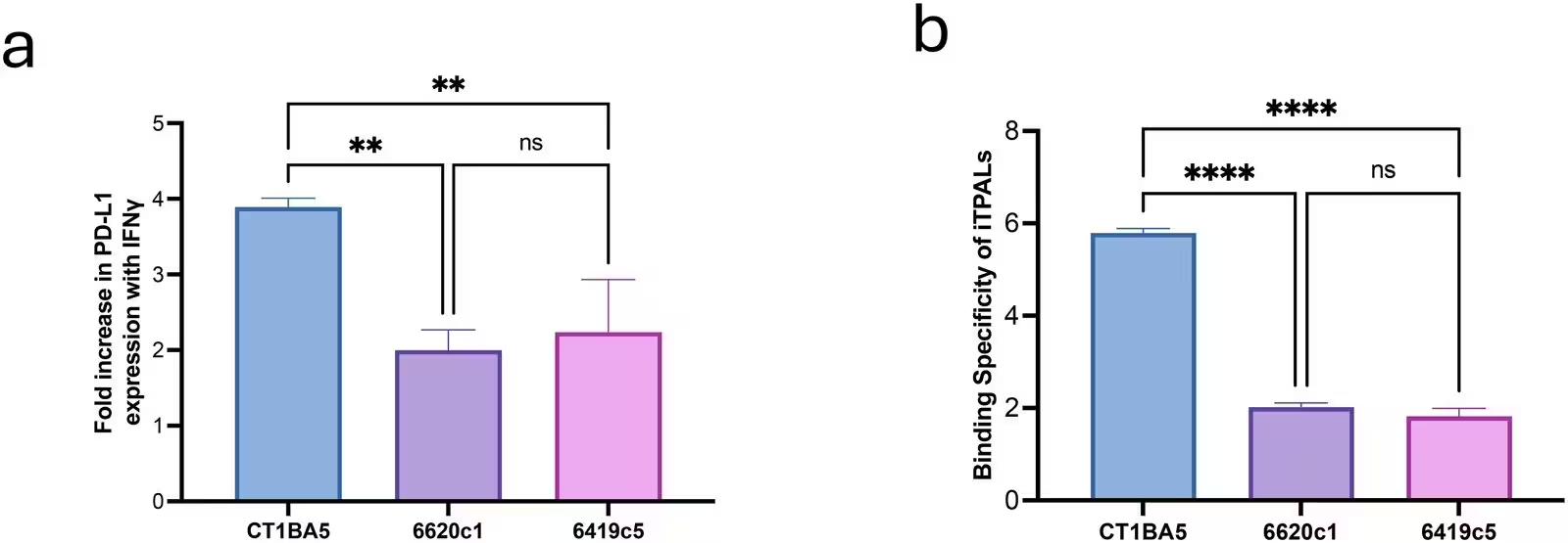
The specialized nanoconstructs, known as PD-L1 immune checkpoint-targeted photoactivatable liposomes or iTPALs, integrate a photosensitizer-lipid conjugate with surface-conjugated antibodies designed to bind specifically to the PD-L1 protein on cancer cells. PD-L1 is a molecular shield that tumors frequently use to evade detection by the immune system. When exposed to light at a wavelength of 690 nanometers, these liposomes generate reactive oxygen species and stress responses that help dismantle tumor defenses while simultaneously blocking the inhibitory immune checkpoint.
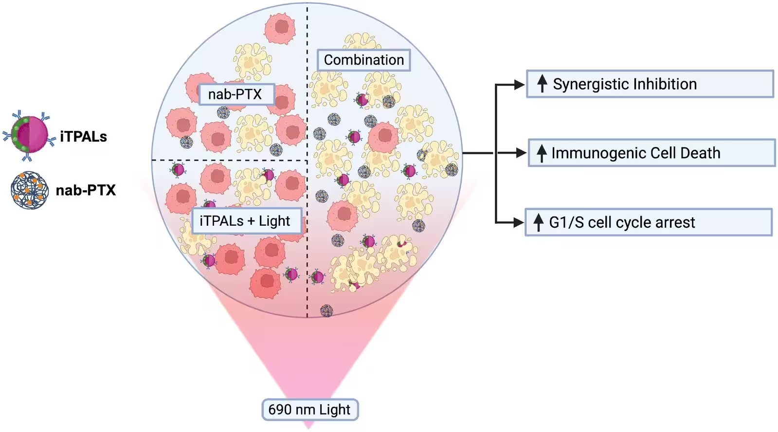
To evaluate the interaction between the two therapies, the team utilized SynergyFinderPlus, a computational tool used to measure drug combinations. The analysis revealed a powerful synergistic inhibition of metabolic activity across all tested cancer cell lines, with mean synergistic inhibition scores exceeding eighty percent regardless of whether the chemotherapy was administered before or after light activation. Importantly, the experiments showed no antagonistic or merely additive effects at any tested dosage combination, confirming that the two treatments work harmoniously together.
Does the Combination Alter Cellular Uptake and Cycle Progression?
Although previous scientific literature suggested that photodynamic therapy might enhance the cellular uptake of chemotherapeutic agents, liquid chromatography-mass spectrometry measurements revealed no significant changes in intracellular paclitaxel concentrations when combined with light-activated iTPALs at the tested doses. Instead, the synergy appears to stem from complementary mechanisms involving cell cycle regulation and immune signaling pathways. Standard paclitaxel typically stabilizes microtubules and arrests cells at the mitosis phase, whereas light-activated iTPALs induced pronounced cell cycle arrest at the earlier G1/S transition phase.
Furthermore, the team assessed immunogenic cell death, a specialized form of cell death that alerts the immune system to the presence of danger through the surface exposure of damage-associated molecular patterns. While individual treatments showed variable results depending on the specific cell line’s baseline sensitivity, the combination treatment significantly amplified the surface exposure of key stress markers such as calreticulin, HSP70, and HSP60. Notably, this enhancement overcame the lack of strong immunogenic signaling typically observed in certain resistant cell lines when treated with chemotherapy alone.
What Are the Next Steps for This Photodynamic Chemoimmunotherapy?
While the findings demonstrate high synergistic efficacy in laboratory cell cultures, the authors emphasize that these results serve as an in vitro proof of concept. Because the experiments were conducted using isolated cell lines, future investigations will need to determine whether the observed immunogenic enhancements translate into successful tumor regression and survival extensions in living animal models with intact immune systems. Subsequent studies will also explore how iTPAL-mediated tissue remodeling influences drug penetration in complex living tumor environments, bringing researchers one step closer to more effective clinical strategies for hard-to-treat cancers.
The research was published in RSC pharmaceutics on November 19, 2026.
This article has been fact checked for accuracy, with information verified against reputable sources. Learn more about us and our editorial process.
Last reviewed on .
Article history
- Latest version
Reference(s)
- Bhandari, Chanda., et al. “Synergistic enhancement of in vitro nab-paclitaxel responses by PD-L1 immune checkpoint targeted photoactivable liposomes with altered cell-cycle progression and amplified immunogenic signaling.” RSC Pharmaceutics, January 1, 2027 Royal Society of Chemistry (RSC), doi: 10.1039/d6pm00480f. <https://doi.org/10.1039/d6pm00480f>.
Cite this page:
- Posted by Robert Brown