Light-Powered Nanorobots Swim Through Bloodvessels To Rescue Damaged Heart Muscle Cells
Engineered biohybrid motors restore cellular energy and reverse tissue scarring in animal models of chemotherapy-induced heart damage.
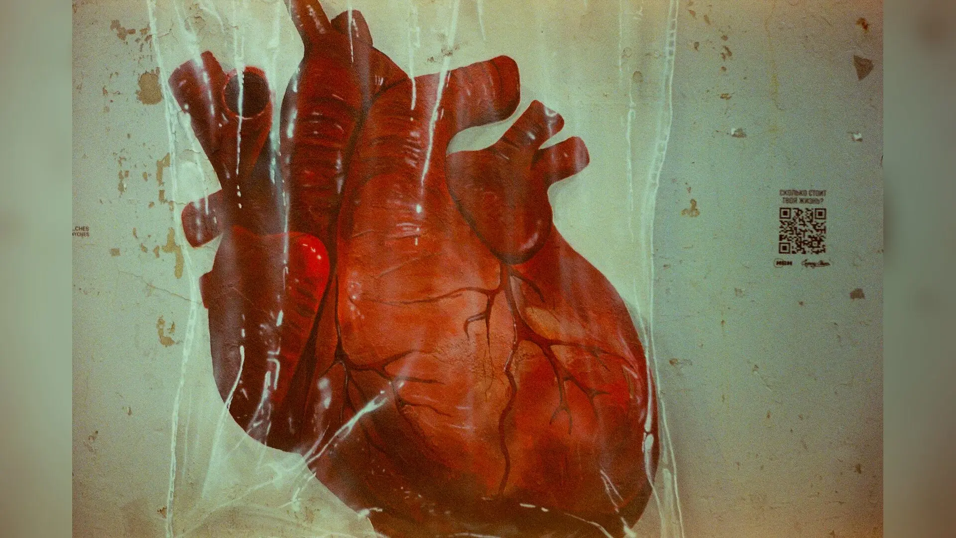
Heart muscle cells damaged by chemotherapy can now be rescued using light-activated nanorobots that swim through the bloodstream to deliver fresh cellular energy.
Myocardial injury, often triggered by cardiotoxic cancer treatments like doxorubicin, leads to a severe energy crisis inside cells. When the microscopic power generators known as mitochondria fail, heart tissue suffers from a shortage of cellular fuel, damaging chemical imbalances, and scarring that can lead to heart failure.
How Do Light-Driven Nanorobots Restore Heart Muscle Cell Energy?
To tackle this energy deficit, a research team led by Yue and colleagues designed a self-directed biohybrid nanorobot called an ATPNbot. These microscopic machines feature an asymmetric, two-sided structure built from natural photosynthetic bacterial membranes on one half and a synthetic lipid shell on the other. When exposed to structured near-infrared light, which can penetrate deep into tissue, the nanorobot acts as a miniature power generator.
The photosynthetic components capture the light energy to drive rotary protein motors that synthesize fresh adenosine triphosphate, the primary energy currency of living cells, directly from internal building blocks. This catalytic action propels the nanorobots forward, allowing them to navigate along light gradients toward damaged areas. The study detailing these findings was published in Science Advances.
Can Microscopic Machines Navigate Biological Barriers in Living Organisms?
Successfully treating heart tissue requires navigating a complex gauntlet of biological barriers, including the cell-lining layers of blood vessels and dense cardiac tissue. Laboratory tests showed that the nanorobots utilize two entry pathways, primarily macropinocytosis, to enter heart muscle cells. Once inside, they use light activation to escape from destructive digestive compartments known as lysosomes and disperse throughout the cytoplasm.
When tested in models of doxorubicin-induced cardiotoxicity, the ATPNbots rapidly increased intracellular energy levels to roughly double baseline values. This influx of energy helped restore the chemical balance of oxidized and reduced nicotinamide adenine dinucleotide coenzymes, stabilized mitochondrial membrane potential, and reduced harmful reactive oxygen species by about half. Furthermore, the treatment significantly suppressed destructive pore opening in mitochondria and smoothed out erratic calcium oscillations in injured cells.
What Improvements Occur in Animal Models After Treatment?
In laboratory mice with drug-induced heart damage, intravenous administration of the nanorobots guided by near-infrared light led to a two-point-four-fold higher accumulation in heart tissue compared to passive control vesicles. Treated mice maintained stable body weights and showed significantly improved survival rates.
Echocardiography demonstrated that the therapy restored left ventricular ejection fraction and fractional shortening toward healthy baseline levels. Histological analysis revealed that the treatment reduced collagen deposition and scarring to less than six percent of the tissue area, while preserving normal muscle fiber structure and preventing the cellular shrinkage seen in untreated injury models.
While extensive preclinical safety studies and further trials are necessary before these systems can be considered for human patients, the work establishes a foundation for active, light-guided energy medicine that goes beyond traditional passive drug delivery.
The research was published in Science advances on November 20, 2026.
This article has been fact checked for accuracy, with information verified against reputable sources. Learn more about us and our editorial process.
Last reviewed on .
Article history
- Latest version
Cite this page:
- Posted by Robert Brown