Scientists Crack Tough Tendon Barrier to Watch Living Cells Activate Repair Genes
A newly optimized molecular sensor delivery method allows real-time tracking of gene expression inside living tendon tissue following mechanical injury.

Tendon injuries and chronic degeneration affect millions of people worldwide, often leading to lasting pain and mobility loss because current diagnostic tools destroy the very tissue they aim to study. Researchers have long struggled to watch living tendon cells activate specific genes over time, blocked by a dense, fibrous collagen shield that keeps out molecular probes. A team of scientists led by Mona Ahmed, Daniel Robles, Ninghao Zhu, Spencer Szczesny, and Pak Kin Wong has now cracked this barrier by engineering a prolonged delivery method for specialized molecular sensors into intact tendon fascicles, which are small bundles of tendon fibers.
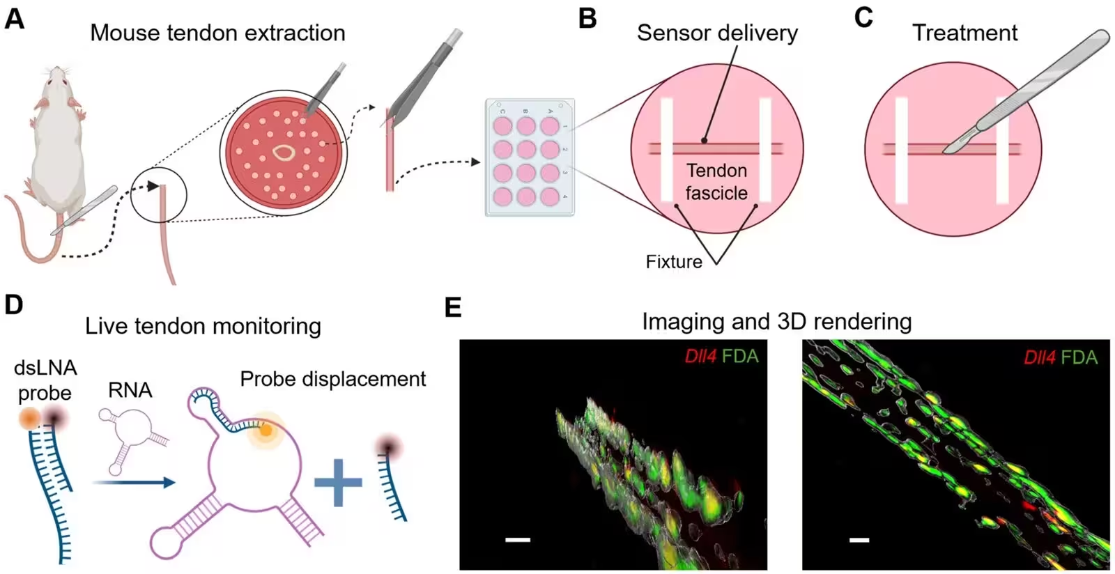
The study, published in The Analyst, details a technique using double-stranded locked nucleic acid biosensors, which are fluorescent probes paired with a quencher molecule. When the sensor encounters its specific target messenger RNA, which carries genetic instructions from DNA to protein-building machinery, it releases the quencher and lights up. While standard delivery protocols fail inside tough tendon matrix, extending the incubation period with a cationic lipid carrier to five days successfully transports the sensors into viable tenocytes, the specialized cells that maintain tendon tissue, without killing them.
How Living Tendon Fascicles Reveal Genetic Signals After Injury
To demonstrate the platform, the authors targeted a gene called Delta-like ligand 4, abbreviated as Dll4, which encodes a key membrane-bound protein involved in cellular communication and wound healing. In many tissues, physical injury disrupts local mechanical tension, triggering Dll4 expression to guide the formation of specialized leader cells that direct tissue repair. The team isolated tail tendon fascicles from mice, secured them in imaging plates, and created controlled mechanical wounds using a surgical blade while tracking fluorescence via confocal microscopy over several days.
Within 24 hours of injury, the Dll4 biosensor signal surged dramatically right along the wound edge compared to the quiet tendon body far from the cut. This spatial concentration points to a localized genetic response triggered by mechanical disruption. When the researchers blocked a critical cellular pathway called gamma-secretase using a chemical inhibitor named DAPT, the wound-edge Dll4 signal dropped significantly. Inhibiting a different regulatory protein called ROCK using a compound designated Y-27632 altered the spatial distribution of Dll4, driving up signals across the broader tendon body while shifting how cells responded at the injury boundary.
What These Cellular Changes Mean for Future Tendon Therapies
The ability to track gene activity in real time inside living 3D tendon tissue bridges a major gap between simple two-dimensional cell dishes and complex whole-animal models. Conventional methods like RNA sequencing or PCR require destroying the tissue, making it impossible to watch a single injury heal day by day. By demonstrating that lipid-based carriers can penetrate dense collagen matrices given enough time, the work opens new avenues for studying tendinopathy, overuse injuries, and tissue degeneration.
The authors acknowledge important limitations in their work, noting that fluorescence intensity currently serves as a relative, semi-quantitative measure rather than an absolute count of mRNA molecules. Furthermore, the long-term impact of extended biosensor incubation on normal cell behavior requires deeper investigation, and the exact functional role of Dll4 signaling in tendon repair remains to be fully mapped. Future refinements to probe chemistry and delivery systems could soon allow scientists to monitor multiple disease-related pathways simultaneously under dynamic mechanical loading.
The research was published in The Analyst on November 17, 2026.
This article has been fact checked for accuracy, with information verified against reputable sources. Learn more about us and our editorial process.
Last reviewed on .
Article history
- Latest version
Reference(s)
- Ahmed, Mona., et al. “A locked nucleic acid biosensor for gene expression analysis in live tendon tissue.” The Analyst, January 1, 2026 Royal Society of Chemistry (RSC), doi: 10.1039/d6an01121g. <https://doi.org/10.1039/d6an01121g>.
Cite this page:
- Posted by Rohan Kumar