Brain Stimulation Triggers Artificial Hibernation That Protects Rat Hearts From Oxygen Deprivation
Activating specific hypothalamic neurons induces a synthetic torpor state that shields cardiac tissue from severe injury, even without body cooling.
Rats forced into a temporary, artificial hibernation state experience a stunning 40 percent drop in heart tissue damage when deprived of oxygen, according to a recent study. Researchers found that triggering specific neurons in the brain can shield the heart from severe injury without even needing the animal’s body temperature to drop.
During times of environmental stress, many animals enter torpor, which is a reversible protective physiological state typically characterized by reductions in core temperature, heart rate, and oxygen consumption. Species that naturally enter this hypothermic and hypometabolic state are remarkably tolerant of ischaemia-reperfusion injury, a type of tissue damage that occurs when blood supply returns to tissue after a period of oxygen deprivation. Consequently, there is a growing interest in utilizing aspects of torpor for clinical applications, such as protection from stroke or myocardial infarction. It was previously unknown, however, whether a torpor-like state could protect animals that do not naturally enter torpor.
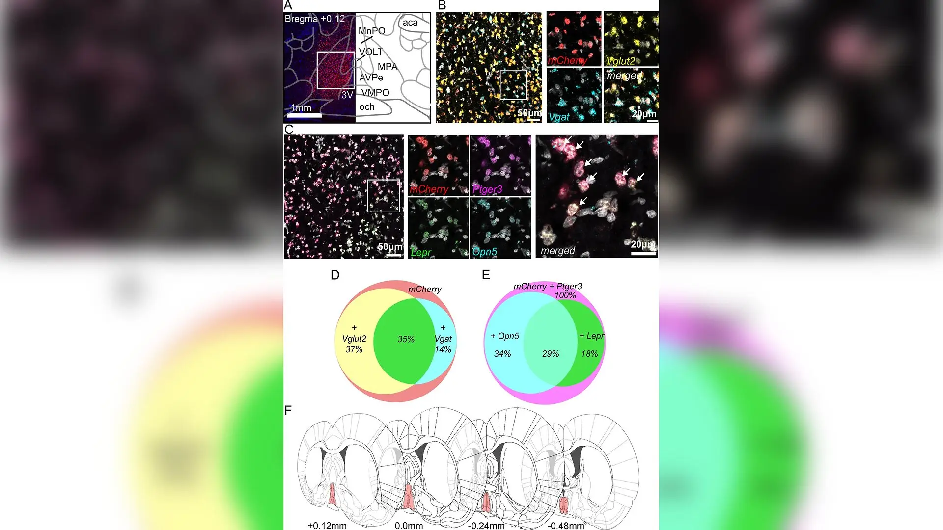
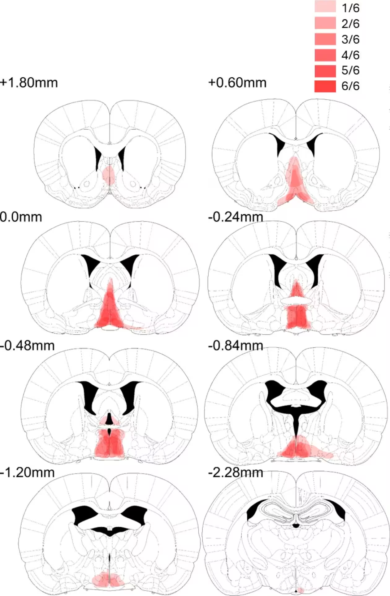
To investigate this, a team of researchers led by scientists at the University of Bristol published in eLife turned to the rat, a species that does not naturally exhibit torpor. Using viral vector-mediated chemogenetics, the team selectively targeted a population of neurons in the medial preoptic area of the hypothalamus, a brain region involved in regulating body temperature and metabolism. By delivering a modified receptor into these specific brain cells, the researchers were able to activate them using a synthetic drug called clozapine-N-oxide.
How Brain Stimulation Mimics Natural Hibernation
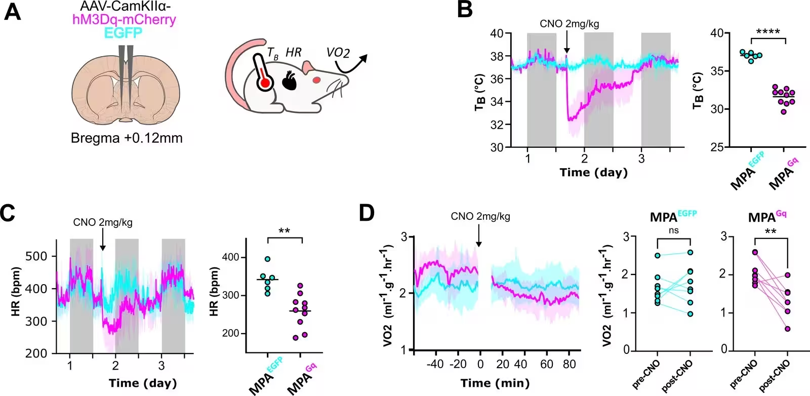
Activating these targeted neurons in the rat hypothalamus successfully induced a synthetic torpor state. The treated animals experienced a rapid and reversible decrease in core body temperature, dropping from a normal average of about 37 degrees Celsius down to roughly 31.6 degrees Celsius. This was accompanied by a 24 percent reduction in heart rate and a 39 percent decrease in overall oxygen consumption, successfully mimicking the key physiological hallmarks of natural torpor.
The researchers then tested whether this centrally induced state could protect cardiac tissue against simulated heart attacks. Hearts were removed from the rats and studied in an ex vivo model, where they were intentionally deprived of oxygen for 30 minutes before having their flow restored. Pre-conditioning the rats with 90 minutes of synthetic torpor prior to heart removal resulted in a dramatic 38.8 percent reduction in infarct size, meaning the area of dead heart tissue shrank significantly compared to control animals.
Why Body Cooling Was Not Required for Protection
A major question in hibernation research is whether organ protection is merely a side effect of the cold body temperature, or if the brain actively triggers cellular defense mechanisms. To find out, the team housed a group of modified rats at a thermoneutral ambient temperature of 28 to 30 degrees Celsius, which completely prevented the drop in core body temperature when the synthetic torpor drug was administered.
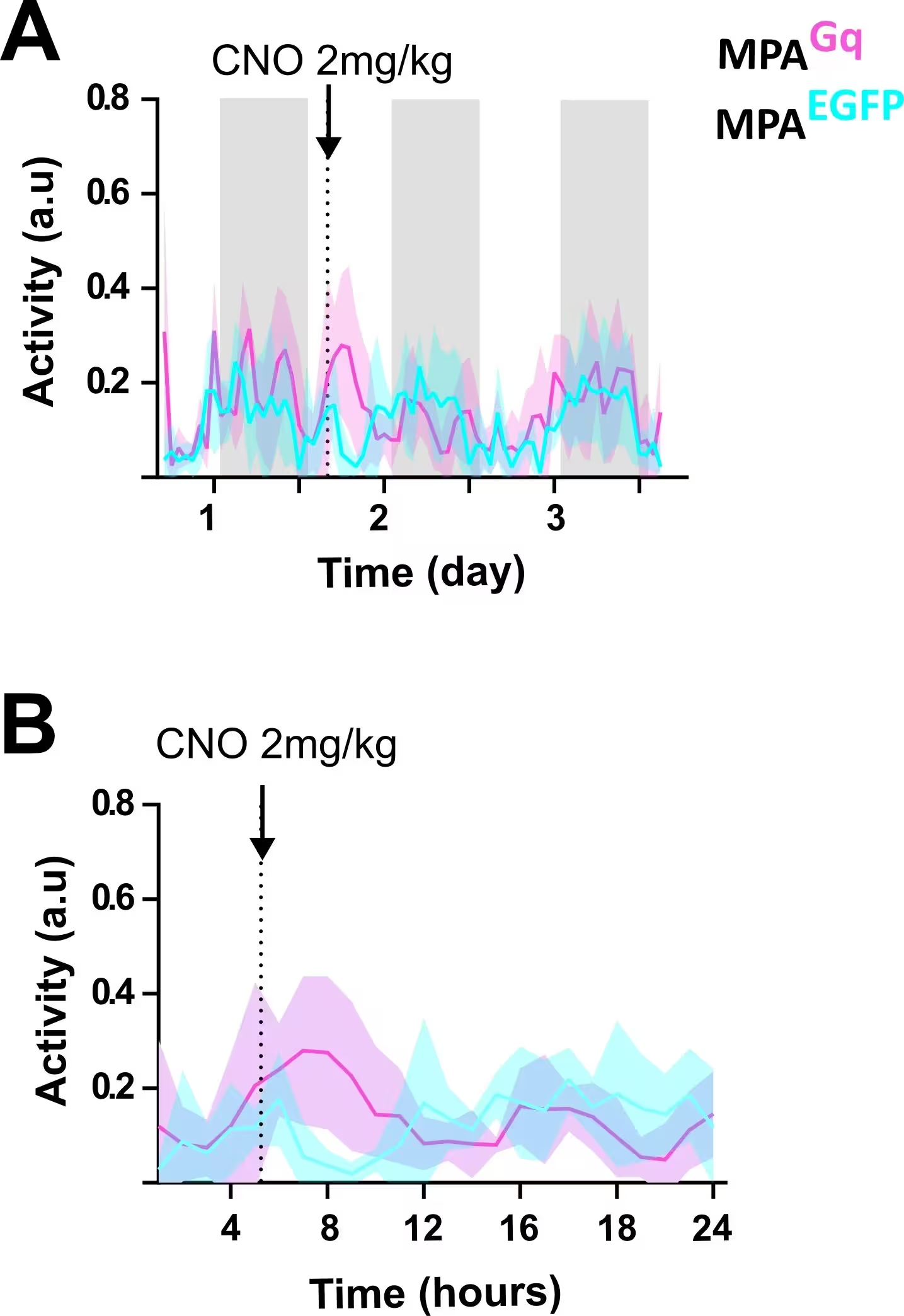
Astonishingly, even when the rats were kept completely warm during the brain stimulation, their isolated hearts still showed a profound reduction in tissue damage when exposed to oxygen deprivation. The data revealed that the myocardial protection conferred by synthetic torpor is independent of hypothermia. Although the animals still experienced a slower heart rate, or bradycardia, this heart rate slowing did not persist once the hearts were isolated in the laboratory system, confirming that simple physical cooling was not driving the protective effect.
Cellular Pathways Revealed by Phosphoproteome Analysis
To uncover the molecular changes happening inside the heart cells, the investigators conducted advanced mass spectrometry to analyze the cardiac phosphoproteome, mapping thousands of protein phosphorylation sites. The analysis showed a coordinated ensemble of pathway modulation, including the suppression of mTOR signaling, which reduces cellular growth drive and anabolism, much like what is observed during calorie restriction or natural hibernation. Suppressing this pathway is known to limit cell death and regulate protective autophagy.
The researchers also observed dampened stress-activated MAPK pathways and altered calcium-handling kinases such as CAMKII, which help maintain heart rhythm and prevent destructive calcium overload during oxygen reperfusion. In parallel, they found increased activity of CSNK2A2, a protein kinase associated with promoting cell survival, DNA repair, and inhibition of apoptosis. Together, these findings indicate that activating specific neurons in the brain commands the heart to switch on a robust, multi-layered survival program.
While this research is still in early-stage laboratory testing and any potential translation to human medicine remains a long way off, the discovery opens exciting new avenues for protecting organs during major surgeries, organ transplants, or heart attacks. Future investigations will need to determine whether synthetic torpor can protect organs when applied after an injury has already begun, and precisely how the brain signals the heart to activate these powerful defense pathways.
The research was published in eLife on November 12, 2026.
This article has been fact checked for accuracy, with information verified against reputable sources. Learn more about us and our editorial process.
Last reviewed on .
Article history
- Latest version
Reference(s)
- Elley, Megan., et al. “Synthetic torpor in the rat protects the heart from ischaemia-reperfusion injury.” eLife, vol. 15, November 12, 2026 eLife Sciences Publications, Ltd, doi: 10.7554/eLife.110559. <https://doi.org/10.7554/eLife.110559>.
Cite this page:
- Posted by Priya Sharma