Aggressive Liver Cancer Cells Leave A Unique Chemical Fingerprint
Nuclear magnetic resonance profiling reveals that virulent hepatocellular carcinoma cells secrete distinct amino acid derivatives and organic acids into their environment.
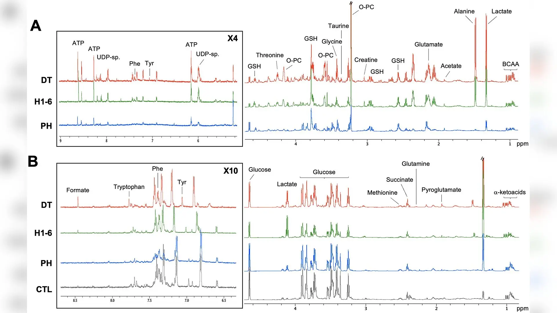
Aggressive liver cancer cells secrete distinct chemical signatures into their surroundings, offering a fresh window into how malignant tumors outpace healthy tissue. Researchers tracked these metabolic shifts to understand why certain cancer strains grow so rapidly.
What is hepatocellular carcinoma and how do normal liver cells compare to tumors?
Hepatocellular carcinoma is the most common form of primary liver cancer and remains a leading cause of cancer mortality worldwide. Treating this disease is notoriously difficult due to high recurrence rates, aggressive metastasis, and resistance to standard therapies. A team of scientists led by Corentine Goossens and colleagues set out to map the hidden biochemical differences that drive this aggressive behavior by studying the internal and external chemical environments of liver cells.
To uncover these patterns, the researchers examined murine primary hepatocytes alongside two liver cancer cell lines, namely the standard Hepa1–6 line and its more aggressive derivative, Dt81Hepa1–6. Using advanced nuclear magnetic resonance spectroscopy, a powerful analytical technique that identifies molecular structures using magnetic fields, the team measured both intracellular metabolites, known as the endometabolome, and extracellular nutrients found in culture media, known as the exometabolome, at 24 and 48 hours. The findings were published in PLOS One, detailing how cancer cells reconfigure their internal machinery to fuel unbridled growth.
Why do cancer cells exhibit the Warburg effect and shift their chemical output?
The analysis revealed a sharp metabolic divide between healthy liver cells and tumor cells. In alignment with the well-known Warburg effect, where cancer cells preferentially rely on glycolysis even when oxygen is abundant, the HCC cell lines displayed markedly higher intracellular and extracellular lactate levels coupled with depleted glucose stores. This accelerated sugar consumption is paired with elevated alanine, pointing to altered pyruvate handling to support rapid cellular proliferation.
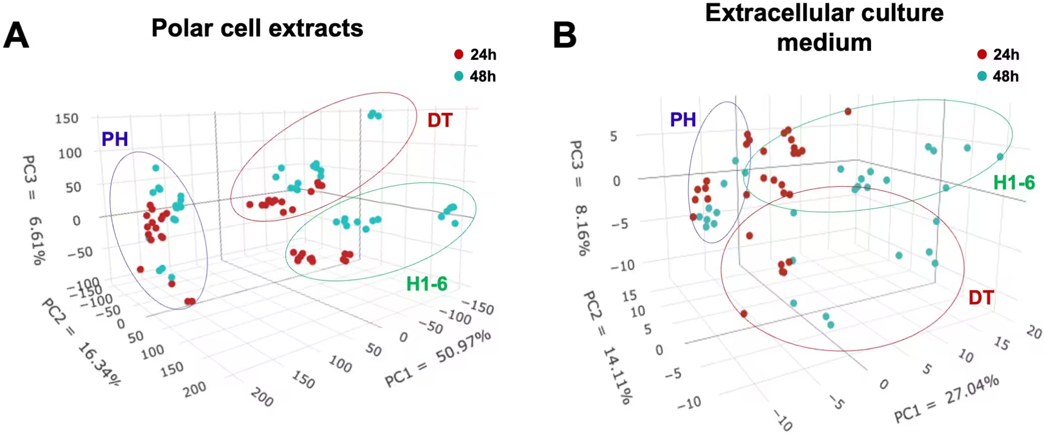
Furthermore, tumor cells showed significant reductions in intracellular glutathione and acetate, signaling a major overhaul in redox balance and central carbon processing. In contrast, compounds linked to osmotic regulation and membrane turnover, such as taurine, creatine, and myo-inositol, were significantly elevated in the cancer lines. These chemical alterations highlight the profound metabolic plasticity required for malignant cells to survive and expand.
What metabolic fingerprints reveal a more aggressive cancer phenotype?
The most striking discoveries emerged when comparing the standard tumor line against its highly aggressive derivative, Dt81Hepa1–6. The aggressive lineage produced a distinct extracellular footprint defined by the significant accumulation of branched-chain amino acid derived ketoacids, specifically 2-oxoisovalerate and 3-methyl-2-oxovalerate. This accumulation indicates profound shifts in nitrogen and carbon handling within the more virulent cells.
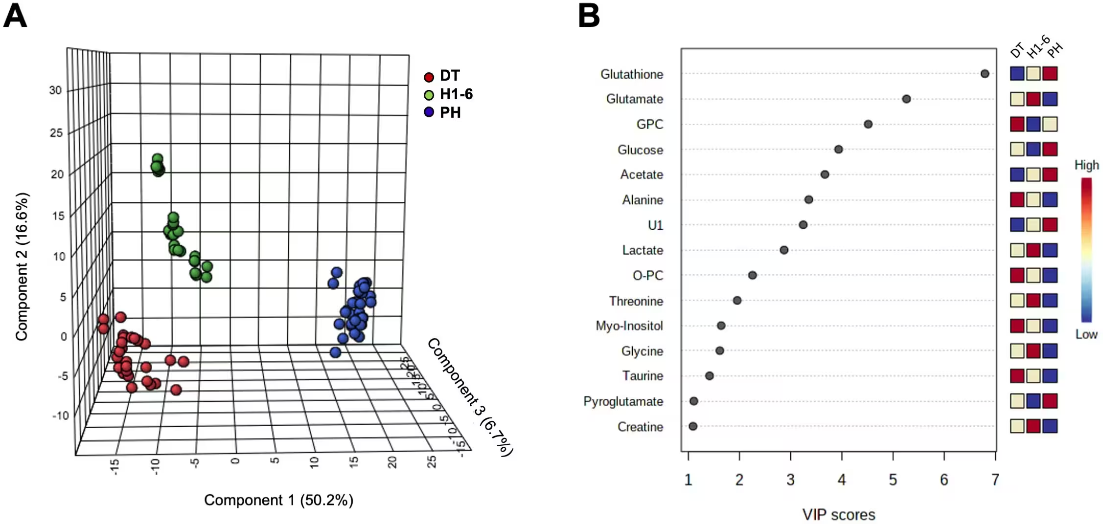
Additionally, the aggressive cells released higher amounts of pyroglutamate, formate, and citrate into their surrounding media. Pyroglutamate is an endogenous molecule tied to glutathione recycling and cellular defense against oxidative stress. Its heightened export, alongside diminished internal glutathione, suggests that highly aggressive liver cancer cells heavily remodel their antioxidant defenses to manage the stress of rapid tumor progression.
What are the primary limitations and future steps for this research?
While the study provides a comprehensive map of metabolic differences, the authors note several limitations. Nuclear magnetic resonance profiling captures relative metabolite abundance but does not directly measure metabolic flux, meaning these findings represent static signatures rather than absolute proof of pathway activity rates. Furthermore, the experiments relied on in vitro mouse cell models, which cannot fully replicate the complex microenvironment and cellular heterogeneity of human liver tumors.
Future investigations will require isotope-tracing experiments and in vivo validations to determine whether these candidate metabolites drive tumor aggressiveness directly or simply reflect downstream byproducts. Extending this dual-compartment metabolomic approach to human patient samples could eventually aid in identifying circulating biomarkers for early liquid biopsy diagnostics.
The research was published in PLOS One on November 12, 2026.
This article has been fact checked for accuracy, with information verified against reputable sources. Learn more about us and our editorial process.
Last reviewed on .
Article history
- Latest version
Reference(s)
- Goossens, Corentine., et al. “NMR-based metabolomic profiling reveals distinct metabolic signatures of primary hepatocytes and hepatocellular carcinoma cell lines.” PLOS One, vol. 21, no. 11, November 12, 2026, pp. e0361879 Public Library of Science (PLoS), doi: 10.1371/journal.pone.0361879. <https://doi.org/10.1371/journal.pone.0361879>.
Cite this page:
- Posted by Hassan Raza