Computational Mapping Reveals Weak Spots in Drug-Resistant Pathogen
Biology

Computational Mapping Reveals Weak Spots in Drug-Resistant Pathogen

A genome-wide screening of 22 bacterial strains suggests older antibiotics could be successfully repurposed against stubborn infections.
By Priya Sharma
Published:
Email this Article
Computational Mapping Reveals Weak Spots in Drug-Resistant Pathogen
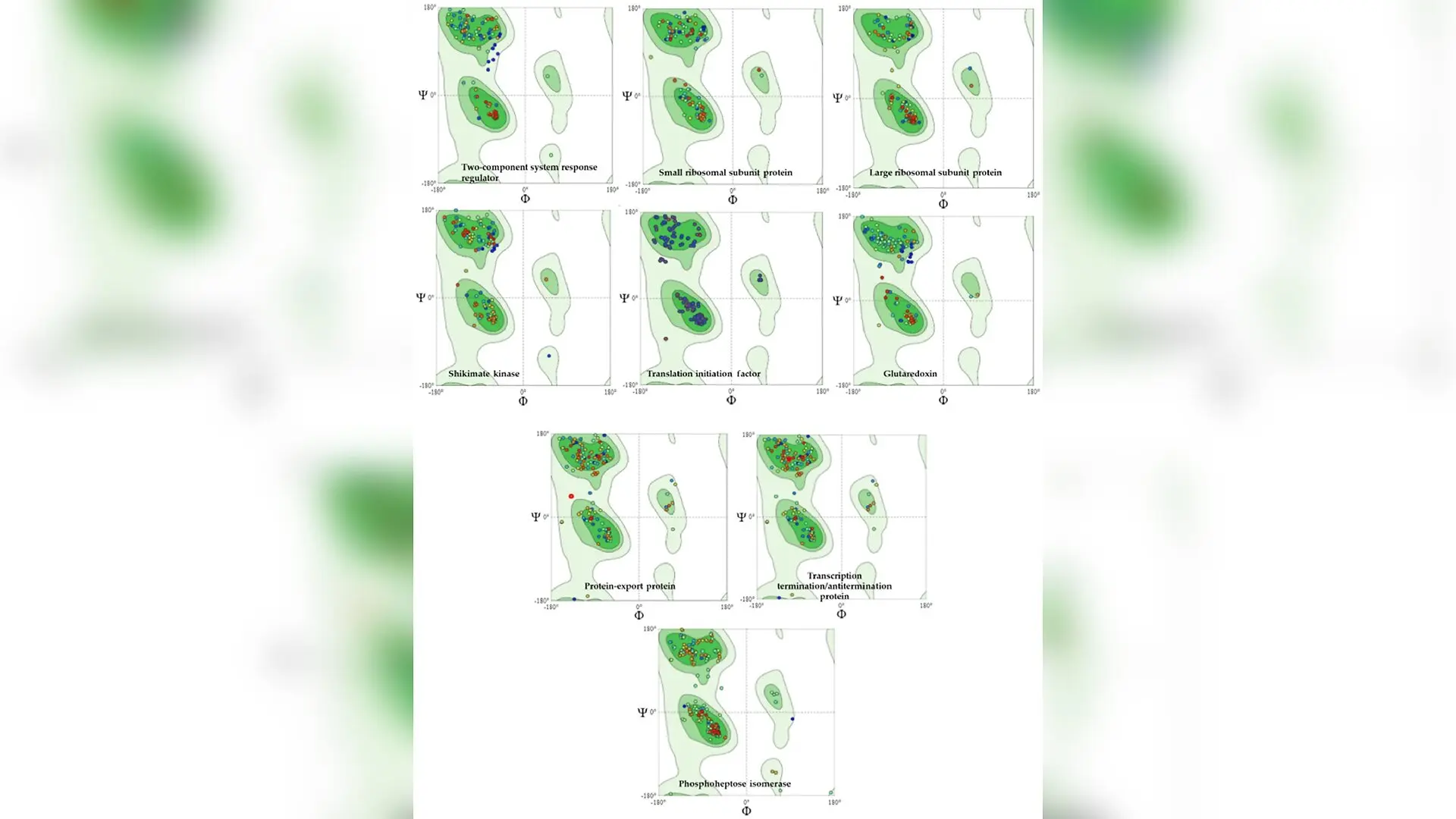
Shows Ramachandran plots for each target protein. The first quadrant, known as quadrant I, represents the region of confirmation in which all residues are permitted. A left-handed alpha band was observed in this region. The region known as quadrant II is the largest in the graph. In particular, the conditions at this location are excellent for atom confirmation. Quadrant III, the largest region following quadrant II, holds this distinction owing to its size. Within this particular geographical area, it has been observed that the prevalence of individuals exhibiting right-handedness is relatively high. This phenomenon is commonly referred to as right-handed alpha. There is hardly any framed location in quadrant IV. The conformation characterized by a torsion angle psi (ψ) ranging from −180 to 0 °and another torsion angle phi (φ) ranging from 0° to 180° is unfavorable, primarily because of steric conflict. Munazza Kanwal et al. / Open medicine (Warsaw, Poland)

The gastrointestinal pathogen Yersinia enterocolitica is increasingly slipping past standard antibiotic defenses, but computational biologists have found potential weak spots by mapping out the bacterium’s entire core proteome. Researchers examined 22 complete genomes of the Gram-negative bacterium, which can cause severe foodborne illness, to figure out how old drugs might be given a second life against resistant strains.

By filtering out proteins that resemble human proteins and focusing strictly on what the pathogen needs to survive, the team narrowed down thousands of genes into a targeted list of essential vulnerabilities. The study, published in Open Medicine, utilized advanced computer modeling to test how 30 different fluoroquinolone antibiotics might bind to these critical bacterial structures.

How Computer Screening Identifies New Drug Targets in Bacteria

Yersinia enterocolitica is an asporogenous, rod-shaped bacterium that often enters the human food supply through contaminated milk or meat, causing symptoms ranging from mild gastroenteritis to pseudoappendicitis. While traditional discovery methods take years of trial and error in wet labs, the research team used subtractive genomics and pan-genome analysis to fast-track the search for new treatments.

Using annotation tools like Prokka and Roary alongside the database EDGAR, the authors identified 3,025 core genes shared across the bacterial strains. Out of these, they isolated 2,267 non-host homologous proteins, meaning they are distinct enough from human proteins to minimize toxic side effects. Further screening against the Database of Essential Genes pointed to 29 proteins absolutely vital for the bacterium to grow and survive.
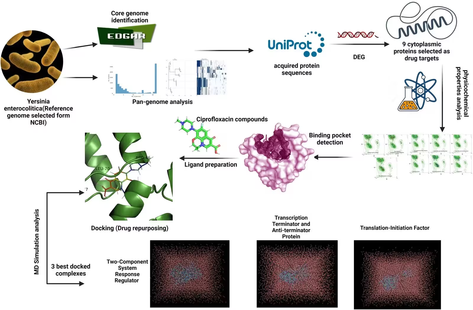
Figure 1: A graphical representation of the computational approach for drug repurposing in Yersinia enterocolitica. Complete genomes were obtained from NCBI and examined to determine the core genome. Subtractive genomics was used to filter core proteins and identify non-host homologous and critical cytoplasmic targets. Prioritized proteins were computationally modeled, screened for druggable binding sites, and docked with fluoroquinolone drugs. The top protein-ligand complexes were validated further using molecular dynamics simulations to determine interaction stability.
A graphical representation of the computational approach for drug repurposing in Yersinia enterocolitica. Complete genomes were obtained from NCBI and examined to determine the core genome. Subtractive genomics was used to filter core proteins and identify non-host homologous and critical cytoplasmic targets. Prioritized proteins were computationally modeled, screened for druggable binding sites, and docked with fluoroquinolone drugs. The top protein-ligand complexes were validated further using molecular dynamics simulations to determine interaction stability. Credit: Munazza Kanwal et al. / Open medicine (Warsaw, Poland)

Which Proteins Showed the Strongest Binding Potential

Out of the essential proteins, nine cytoplasmic targets and one membrane protein were successfully modeled in 3D using the Swiss Model and evaluated for druggable pockets. Molecular docking simulations performed with AutoDock Vina revealed that several older fluoroquinolone drugs, some of which face growing bacterial resistance, still form remarkably stable bonds with specific bacterial machinery.

For instance, the two-component system response regulator OmpR showed strong binding interactions with ciprofloxacin, yielding a docking score of minus 7.4 kilocalories per mole. Meanwhile, the large ribosomal subunit protein uL13 demonstrated the highest overall interaction score of minus 7.8 kilocalories per mole when docked with nalidixic acid. Other promising matches included phosphoheptose isomerase binding with norfloxacin and protein-export protein SecB interacting with ciprofloxacin.
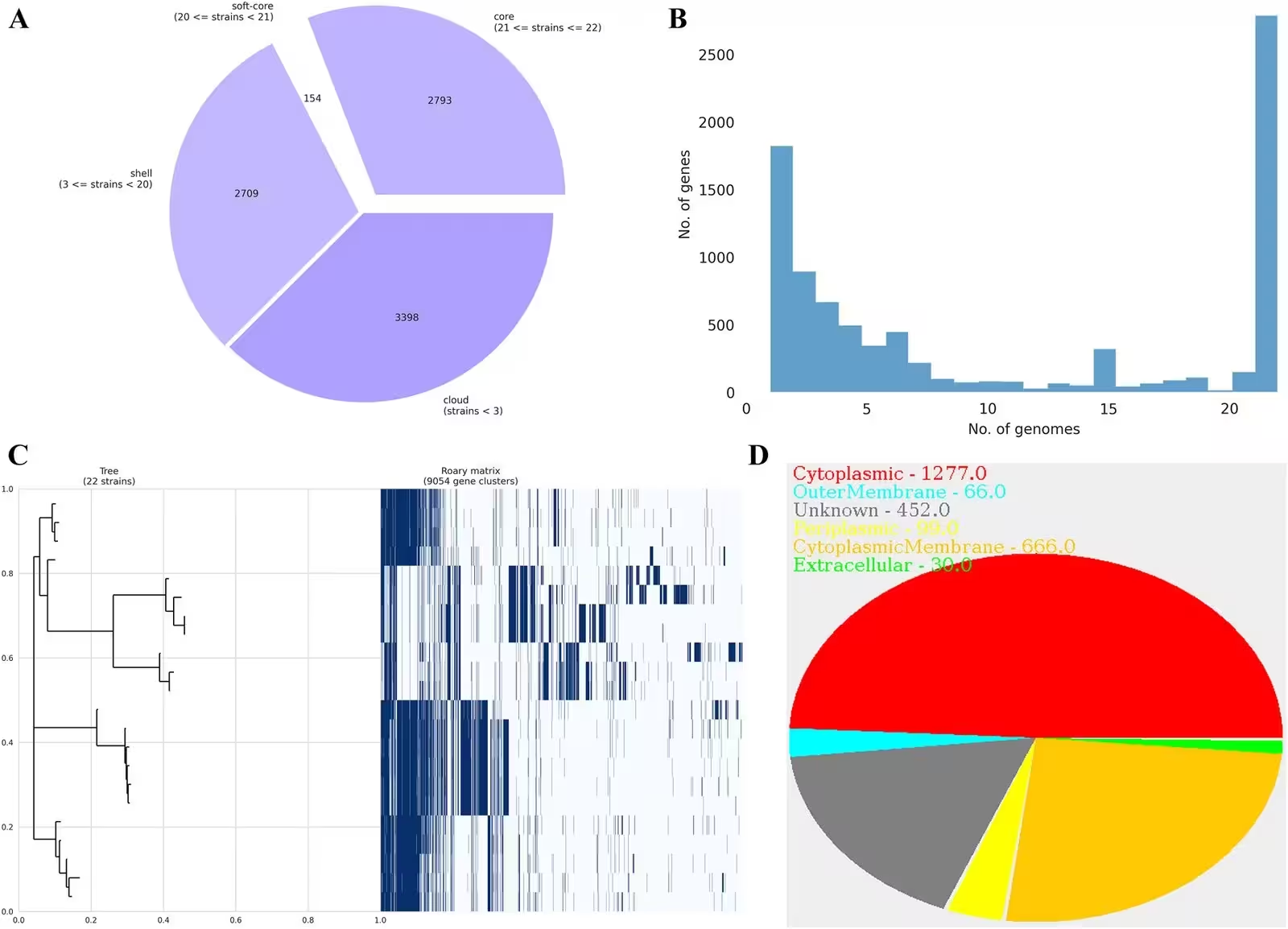
Figure 2: Four panels show gene counts across 22 genomes, gene-family prevalence, a clustered heatmap, and protein subcellular localization. A. The bar graph represents the relationship between the number of genes present in each of the 22 genomes considered. B. The pie chart illustrates the distribution of gene families across 22 strains, categorized into four groups based on their prevalence. The core genome (2,793 genes) includes genes present in all or nearly all strains (21–22 strains). The soft-core genome (154 genes) comprises genes found in 20 strains. The shell genome (2,709 genes) includes genes present in 3–19 strains, while the cloud genome (3,398 genes) represents rare genes found in fewer than 3 strains. This distribution highlights a substantial accessory genome, suggesting high genomic diversity and adaptability within the species. C. The provided image depicts a clustered heatmap integrated with dendrograms, which is a widely employed visualization technique for representing clu
Four panels show gene counts across 22 genomes, gene-family prevalence, a clustered heatmap, and protein subcellular localization. A. The bar graph represents the relationship between the number of genes present in each of the 22 genomes considered. B. The pie chart illustrates the distribution of gene families across 22 strains, categorized into four groups based on their prevalence. The core genome (2,793 genes) includes genes present in all or nearly all strains (21–22 strains). The soft-core genome (154 genes) comprises genes found in 20 strains. The shell genome (2,709 genes) includes genes present in 3–19 strains, while the cloud genome (3,398 genes) represents rare genes found in fewer than 3 strains. This distribution highlights a substantial accessory genome, suggesting high genomic diversity and adaptability within the species. C. The provided image depicts a clustered heatmap integrated with dendrograms, which is a widely employed visualization technique for representing clu Credit: Munazza Kanwal et al. / Open medicine (Warsaw, Poland)

What Molecular Dynamics Simulations Reveal About Drug Stability

To verify that these virtual drug pairings would hold together under physiological conditions, the researchers subjected the top complexes to 100-nanosecond molecular dynamics simulations using the GROMACS software program. Tracking root mean square deviation metrics across the trajectories demonstrated that proteins like OmpR and the transcription terminator maintained stable configurations throughout the simulation runs.

The calculations confirmed that the ligand molecules remained securely anchored within their respective binding pockets without causing structural collapse of the protein backbones. Energy, temperature, and pressure metrics across the simulation boxes further verified system equilibrium, pointing to reliable physical interactions between the repurposed drugs and their targets.

What These Computational Insights Mean for Future Treatments

While these findings rely entirely on in silico modeling rather than clinical trials, they provide a structured roadmap for tackling antimicrobial resistance in hard-to-treat pathogens. By showing that established antibiotics can still latch onto vital internal machinery, the work opens viable pathways for drug repurposing and targeted structural design against Yersinia enterocolitica infections.

The research was published in Open medicine (Warsaw, Poland) on January 1, 2026.

Fact Checked

This article has been fact checked for accuracy, with information verified against reputable sources. Learn more about us and our editorial process.

Last reviewed on .

Article history

  • Latest version

Reference(s)

  1. Kanwal, Munazza., et al. “Molecular insights into drug repurposing and drug target proteins identification for Yersinia enterocolitica.” Open Medicine, vol. 21, no. 1, October 26, 2026 Walter de Gruyter GmbH, doi: 10.1515/med-2026-1547. <https://doi.org/10.1515/med-2026-1547>.

Cite this page:

Sharma, Priya. “Computational Mapping Reveals Weak Spots in Drug-Resistant Pathogen.” BioScience. BioScience ISSN 2521-5760, 26 October 2026. <https://www.bioscience.com.pk/en/subject/biology/molecular-insights-into-drug-repurposing-and-drug-target-proteins-identification-for-yersinia-enterocolitica>. Sharma, P. (2026, October 26). “Computational Mapping Reveals Weak Spots in Drug-Resistant Pathogen.” BioScience. ISSN 2521-5760. Retrieved October 26, 2026 from https://www.bioscience.com.pk/en/subject/biology/molecular-insights-into-drug-repurposing-and-drug-target-proteins-identification-for-yersinia-enterocolitica Sharma, Priya. “Computational Mapping Reveals Weak Spots in Drug-Resistant Pathogen.” BioScience. ISSN 2521-5760. https://www.bioscience.com.pk/en/subject/biology/molecular-insights-into-drug-repurposing-and-drug-target-proteins-identification-for-yersinia-enterocolitica (accessed October 26, 2026).
End of the article