Bone Marrow Protein Loss Shifts Megakaryocyte Positions Without Harming Platelets
Removing a key vascular basement membrane protein alters cell adhesion and spatial organization in mice without disrupting blood clotting.
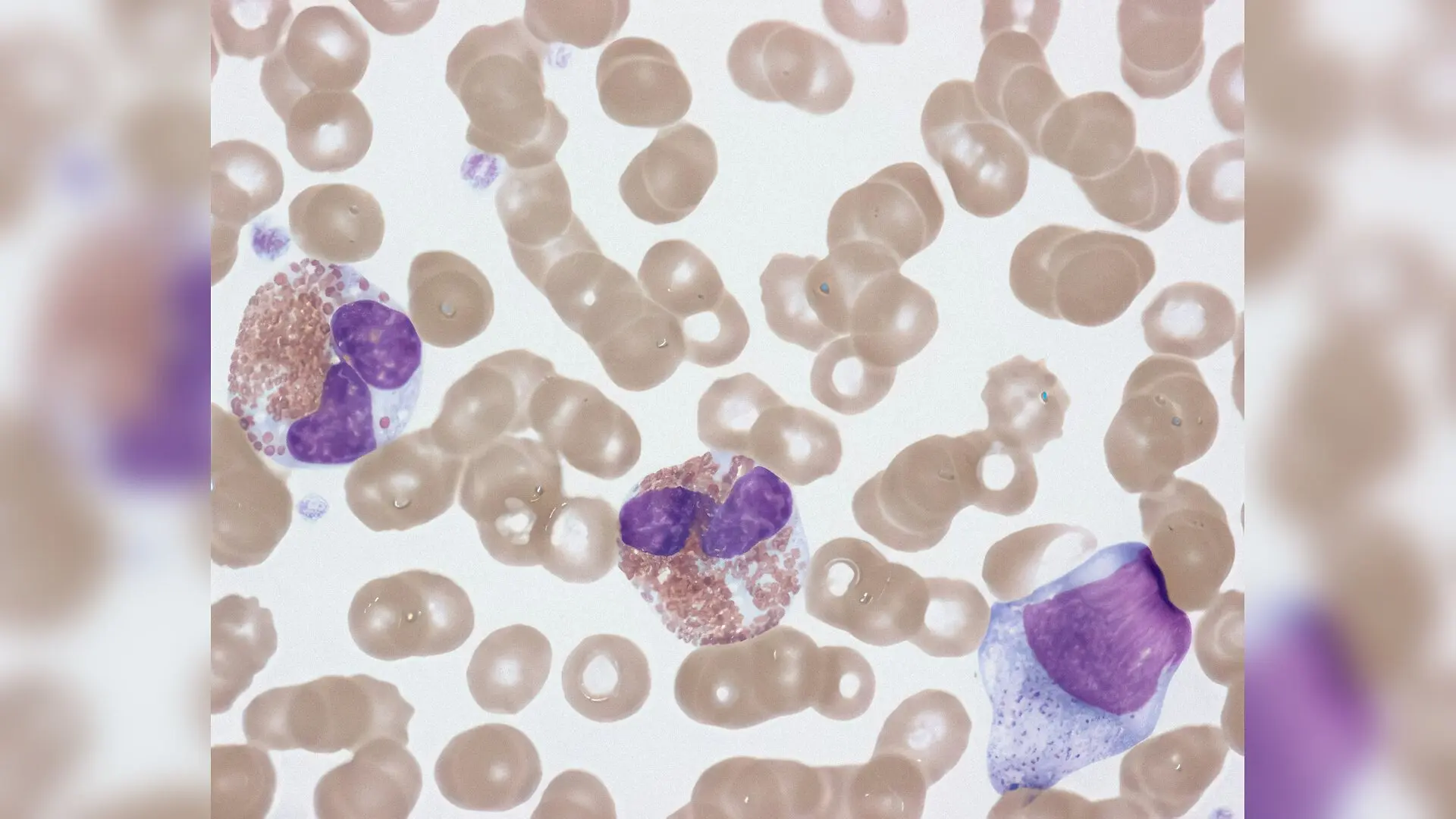
When bone marrow cells lose a key structural protein called laminin alpha 5, blood-producing megakaryocytes shift their position and crowd closer to blood vessels without disrupting overall platelet numbers. The study, published in PLOS One, reveals that this extracellular matrix component acts less like a strict traffic controller for blood production and more like a spatial organizer for the bone marrow niche.
How Platelets Form Inside the Bone Marrow
Platelets are tiny cell fragments essential for blood clotting, and they are continuously manufactured by massive precursor cells residing in the bone marrow known as megakaryocytes. These giant cells extend long cytoplasmic protrusions called proplatelets through the walls of blood vessels, specifically targeting venous sinusoids to release platelets directly into circulation while avoiding leaks inside the bone cavity. Scientists have long suspected that the extracellular matrix, a complex meshwork of proteins surrounding these vessels, guides this cellular choreography.
Among the proteins lining these sinusoidal walls is laminin alpha 5, a large molecule previously thought to play a major regulatory role in governing platelet release. To test this hypothesis, an international team of researchers generated a conditional knockout mouse model where the gene encoding laminin alpha 5 was specifically deleted in vascular endothelial cells. The goal was to observe whether removing this specific protein altered how megakaryocytes interact with the blood vessel wall and whether platelet counts would plummet.
Surprising Normal Blood Counts Despite Molecular Changes
Under normal steady-state conditions, mice lacking laminin alpha 5 displayed entirely normal platelet counts, normal platelet sizes, and completely intact platelet function. Furthermore, the maturation and internal DNA replication cycles of their megakaryocytes proceeded normally. However, a closer look at the molecular profile of these cells revealed notable surface adjustments. Platelets harvested from the mutant animals showed significantly higher surface levels of integrin alpha 6 beta 1, a primary laminin-binding receptor, alongside increased expression of the tetraspanin protein CD9.
Laboratory assays investigating cell behavior further illuminated these changes. When immature bone marrow cells were cultured on specific matrix proteins, megakaryocytes lacking laminin alpha 5 exhibited markedly enhanced adhesion and higher rates of proplatelet formation when exposed to laminin 421 and laminin 511. Despite this increased in vitro activity, the terminal release of functional platelet-like particles remained unaltered, successfully explaining why circulating blood counts remained completely stable in the living animals.
What Happens During Acute Stress and Injury
To determine if laminin alpha 5 becomes critical during high-demand situations, the team subjected the mice to acute hematopoietic stress using antibody-induced platelet depletion and radiation-induced thrombocytopenia. Following rapid platelet destruction, both mutant and wild-type mice mounted robust recovery responses, rebuilding their circulating platelet numbers at comparable speeds. However, microscopic examination of the bone marrow tissue uncovered a striking structural difference in where the megakaryocytes chose to settle.
In normal control mice, megakaryocytes were frequently found dispersed at varying distances from the sinusoidal blood vessels following stress. In contrast, megakaryocytes in the laminin alpha 5-deficient mice accumulated in much higher numbers and huddled in significantly closer physical proximity to the vascular niche. This tighter clustering was likely driven by the upregulation of integrin alpha 6, which enhanced their adhesive grip on alternative laminin isoforms persisting in the basement membrane.
Implications for Understanding Bone Marrow Niches
The authors note that these observations challenge the prevailing view that vascular basement membrane proteins directly dictate the total volume of platelet output. Instead, laminin alpha 5 appears to function primarily as a spatial modulator that fine-tunes how precursor cells position themselves against blood vessel walls. While the study was conducted entirely in murine models and requires cautious translation before applying to human hematopoiesis, it demonstrates that bone marrow architecture can undergo considerable local reorganization without disrupting essential blood-clotting functions.
The research was published in PloS one on November 18, 2026.
This article has been fact checked for accuracy, with information verified against reputable sources. Learn more about us and our editorial process.
Last reviewed on .
Article history
- Latest version
Reference(s)
- Mott, Kristina., et al. “Loss of sinusoidal laminin α5 alters megakaryocyte dynamics but preserves thrombopoiesis.” PLOS One, vol. 21, no. 11, November 18, 2026, pp. e0362188 Public Library of Science (PLoS), doi: 10.1371/journal.pone.0362188. <https://doi.org/10.1371/journal.pone.0362188>.
Cite this page:
- Posted by Priya Sharma