Optical Nanosizer Achieves Sub-Nanometer Precision in 3D Nanoparticle Shape Mapping
Combining linear and radial light polarisation allows standard optical microscopes to resolve the three-dimensional shape and size of nanoparticles with electron-microscope-like precision.
Characterizing the precise dimensions and structural geometry of nanoparticles is essential for applications ranging from targeted drug delivery and medical imaging to environmental monitoring and catalytic manufacturing. While transmission electron microscopy has long served as the industry benchmark for nanoparticle morphometry, its high operating costs, slow throughput, and tendency to damage delicate structures under high-energy electron beams severely limit its utility for routine quality control. To overcome these hurdles, optical techniques have emerged as appealing alternatives because they offer non-destructive analysis across large fields of view using standard microscope hardware.
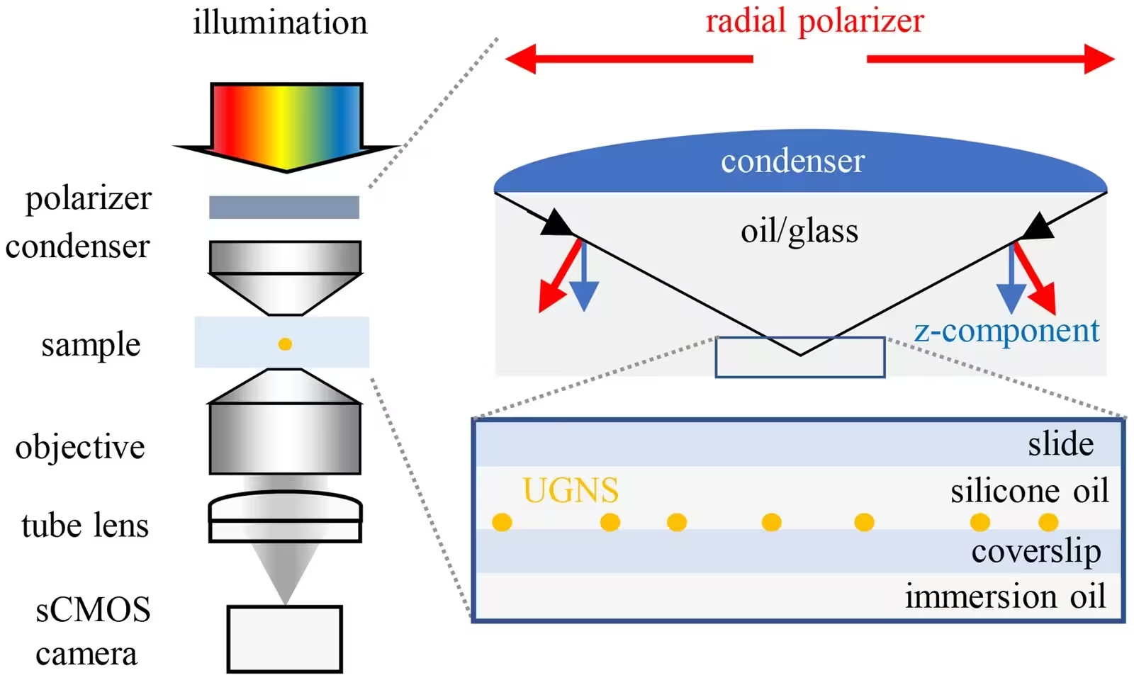
Earlier iterations of the Optical Nanosizer approach successfully mapped in-plane nanoparticle dimensions by capturing wide-field transmission images under varied linear polarizations and wavelengths. However, because standard high numerical aperture condensers focus light primarily within the sample plane, out-of-plane features had to be inferred indirectly through spectral shifts rather than direct measurement. To solve this limitation, the research team introduced measurements incorporating radial polarisation in the condenser back focal plane, generating a robust axial electric field component that directly interrogates the out-of-plane geometry of individual particles. Detailed findings from this investigation are published in Nanoscale, detailing a comprehensive step forward in optical metrology.
Unlocking Three-Dimensional Morphology
The updated optical setup employs both linear and radial polarisation to capture comprehensive scattering and absorption signatures from individual particles. When analyzing ultra-uniform gold nanospheres with a nominal diameter of thirty nanometers, the combined polarization dataset successfully resolves subtle asymmetries and volume metrics that were previously obscured. By comparing experimental extinction cross-sections against theoretical models derived from the Rayleigh-Gans approximation, the system solves the inverse problem to retrieve true three-dimensional particle parameters.
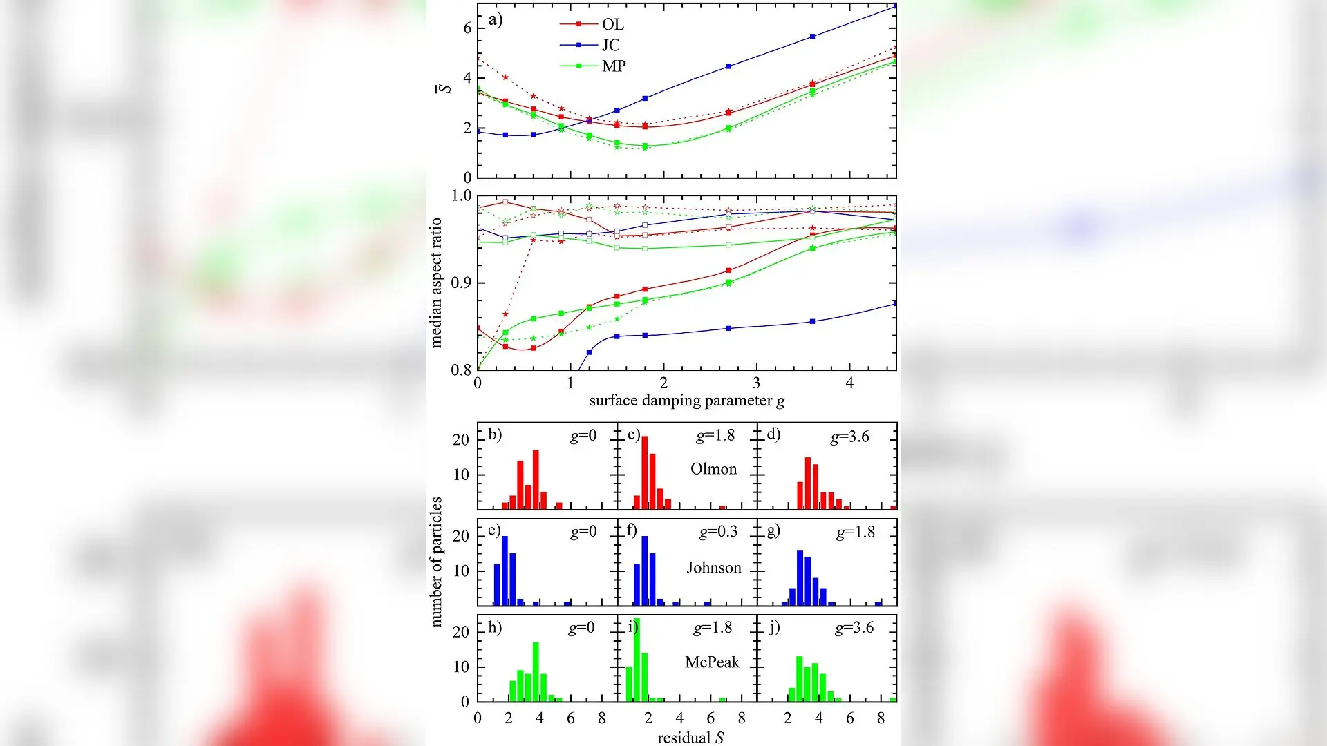
During data processing, the team evaluated several material permittivity datasets from the scientific literature, alongside varying surface damping factors, to determine the most accurate physical description of gold at the nanoscale. Their analysis revealed that incorporating radial polarisation requires careful accounting for optical illumination conditions and system-specific transmission characteristics, such as the long-shadow effect caused by oblique incidence angles. Fine-tuning these parameters significantly improved both the precision and absolute accuracy of size and shape retrievals.
Implications for High-Throughput Metrology
The enhanced Optical Nanosizer demonstrated exceptional performance, pushing size and shape precision below the sub-nanometer threshold for the tested gold nanospheres. While very small particles under twenty nanometers largely avoid retardation effects, particles approaching or exceeding fifty nanometers will eventually require full numerical electrodynamic treatments. Nevertheless, for particles within the size range studied, correcting for minor plasmon resonance red-shifts due to retardation brought optical measurements into remarkable alignment with electron microscopy ground truths.
This optical advancement provides a powerful pathway for high-throughput statistical quality control in nanoparticle production. By bypassing the destructive nature and economic bottlenecks of electron tomography, the enhanced polarization-resolved microscope offers researchers a fast, reliable, and non-invasive tool to map complex nanoscale morphologies across diverse scientific and industrial sectors.
The research was published in Nanoscale on October 22, 2026.
This article has been fact checked for accuracy, with information verified against reputable sources. Learn more about us and our editorial process.
Last reviewed on .
Article history
- Latest version
Reference(s)
- Payne, Lukas M.., et al. “Sub-nanometer 3D morphometric precision of polarisation-resolved wide-field optical extinction microscopy determines the roundness of individual gold nanospheres.” Nanoscale, vol. 18, no. 40, January 1, 2026, pp. 21956-21968. Royal Society of Chemistry (RSC), doi: 10.1039/d6nr03059a. <https://doi.org/10.1039/d6nr03059a>.
Cite this page:
- Posted by John Williams