Structure and Functions of Rabbit Brain
Explore the intricate anatomy and vital functions of the rabbit brain. Learn about its anatomy, functions, and importance in research.
It is the main part of the nervous system. The brain lies in a protective bony box, called cranium. The brain is the anterior part of the central nervous system and lies in the head. It is derived from the embryonic ectoderm. The structural and functional units of the brain are neurons.
(A) Meninges
These are the connective tissue coverings around the brain. The meninges are three. These are the dura mater, arachnoid membrane, and pia mater.
The dura mater is outer and lies close to the inner surface of the cranial cavity. The space between the dura mater and the arachnoid membrane is called the subdural sinus.
The arachnoid membrane is middle and nonvascular. The space between the arachnoid membrane and pia mater is called sub arachnoid sinus. These sinuses are filled with lymph, called cerebrospinal fluid. The fluid and membranes protect the brain from external shocks, prevent desiccation of the brain, and nourish the brain. The pia mater is inner and closely associated with the outer surface of the brain. It is vascular
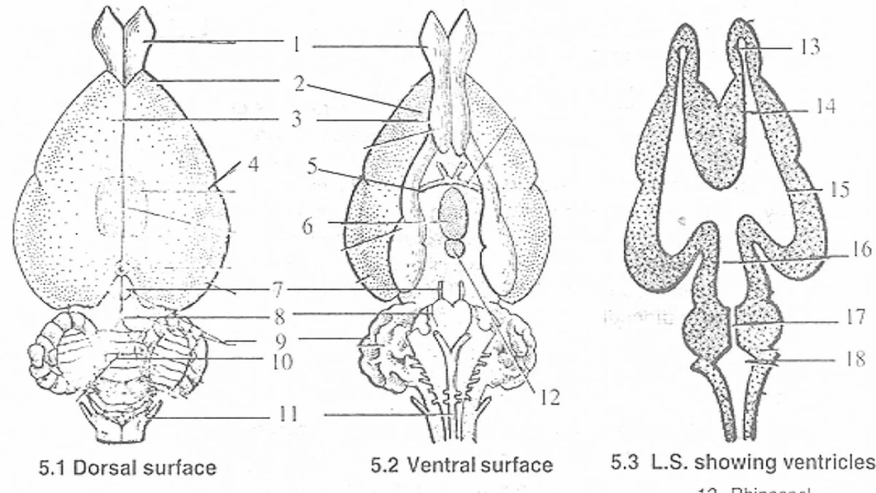
(B) Parts of Brain
The brain is mainly divided into three parts. These are the forebrain, midbrain, and hindbrain.
The forebrain is also called the prosencephalon, it is divided into two parts. These are telencephalon and diencephalon. The telencephalon contains two parts. These are the olfactory lobes and cerebral hemispheres.
Olfactory Lobes
These are a pair, club-shaped, and lie side by side anteriorly. The olfactory lobes are longitudinally separated by groove, called a longitudinal fissure. These are hollow. The cavities of olfactory lobes are called olfactory ventricles or rhinoceros. Each olfactory lobe develops a nerve at its anterior end. This first cranial nerve is called the olfactory nerve.
Cerebral Hemispheres
These two together are called large brains. The cerebral hemispheres are a pair, lying side by side behind the olfactory lobes. Each is half globe-shaped. Each cerebral hemisphere is broader at the posterior end and narrower at the anterior end. These are separated by a longitudinal groove called a cerebral fissure. The surface is smooth without sulci and gyrae. The thin dorsal wall of the cerebral hemisphere is called the pallium. These are also hollow. The cavities are called cerebrocoels or lateral ventricles or '1' and '2' ventricles or paracoels. The cerebral hemispheres contain thick ventrolateral walls. These walls are called corpora striata. The thick ventrolateral walls contain many centers, nerve ganglia, and nerve tracts. The corpora striata are connected by transverse fibers, called anterior commissures. These fibers lie on the floor of paracoels. The roof of the paracoels contains fibers, called corpus callosum. It is the characteristic feature of a mammal's brain. Each cerebral hemisphere contains a central groove in the lateral surface. This groove is called Sylvian fissure. The part of the cerebral hemisphere anterior to the sylvian fissure is called the frontal lobe and the posterior part is called the temporal lobe.
Paracoels anteriorly open into olfactory ventricles and posteriorly into the third ventricle through a common aperture, called the foramen of Monro.
Diencephalon
It is an unpaired part of fore brain. The diencephalon is diamond-shaped. It is anteriorly covered by the posterior ends of the cerebral hemispheres. The dorsal wall contains a pineal stalk and anterior choroid phlexus. The pineal stalk contains a pineal gland. The anterior choroid phlexus is net-like. It is non nervous blood capillary net-like structure. The lateral walls of the diencephalon are thick. These thick walls are called optic thalami. Hence diencephalon is also called thalamencephalon. The ventral wall diencephalon is called the hypothalamus. It contains thirsty and hunger centers. The ventral wall contains another stalk, called the infundibulum. This infundibulum contains an endocrinal gland, called the pituitary. The diencephalon is also hollow. The cavity is called the third ventricle or diocoel.
Mid Brian
It is also called mesencephalon. The midbrain contains four optic lobes. These four optic lobes together are called corpora quadrigemina. It is the characteristic feature of a mammal's brain. The optic lobes are hollow. The cavities are called optocoels. These open into iter or aqueducts Sylvius. The ventral walls of optic lobes are thick, called crura cerebri. The crura cerebri are connected by transverse nerve fibers called posterior commissure.
The optic nerves or second pair of cranial nerves are developed from cruracerebri. They cross one another and form an 'X' shaped optic chiasma on the ventral surface of the diencephalon.
Hind Brain
It is also called rhombencephalon. The hindbrain contains two parts. These are the cerebellum and medulla oblongata. These are unpaired.
Cerebellum
It is the unpaired part. It lies only dorsally. The cerebellum is commonly called the small brain. It contains three parts. The central part is called Vermes. The two lateral lobes are called flocculi. The cerebellum is solid and lies transversely.
Medulla Oblongata
It is the posterior part of the brain. The Medulla oblongata is the most important part of the brain. It is hollow. The cavity is called the fourth ventricle myelocytes or medullary coel. The third and fourth ventricles are connected by a tube called iter or aqueduct of Sylvius. The fourth ventricle posteriorly opens into the central canal of the spinal cord. The medulla posteriorly continues as the spinal cord. The dorsal wall of the medulla contains a non-nervous blood capillary net called posterior choroid phelxus.
Functions of Brain
The brain is the center for the control and coordination of all vital activities. It receives impulses from different parts of the body through sensory nerves and sends orders through motor nerves to different parts of the body for necessary action.
The functions of various parts of the brain are given below:
- The olfactory lobes are the centers of the sense of smell.
- The cerebral hemispheres are the centers of consciousness, intelligence, memory, and voluntary actions.
- The Diencephalon of the brain controls the metabolism of fats and carbohydrates. It also regulates genital functions and sleep. It plays a role in perceiving the sense of vision. Removal of this part of the brain causes blindness.
- Optic lobes are concerned with the sense of sight. They also control the movement of eye muscles.
- The cerebellum serves to coordinate muscular actions and maintain balance during movement.
- The Medulla oblongata is an important part of the brain. It controls involuntary functions such as heartbeat, respiration, excretion, taste, hearing, and sound production. Removal of the medulla oblongata causes the death of the animal.
The information on this page is peer reviewed by a qualified editorial review board member. Learn more about us and our editorial process.
Last reviewed on .
Article history
- Latest version
Cite this page:
- Comment
- Posted by Dayyal Dungrela