Reproductive System of Rabbit (Female)
0
Zoology
Reproductive System of Rabbit (Female)
Explore the female rabbit's reproductive system – anatomy, cycle, and breeding tips. Unlock the secrets of successful rabbit care and breeding.
Published:
The reproductive system of the female rabbit, scientifically known as the doe, is a marvel of nature's engineering. It is a complex and finely tuned system that plays a pivotal role in the perpetuation of this species.
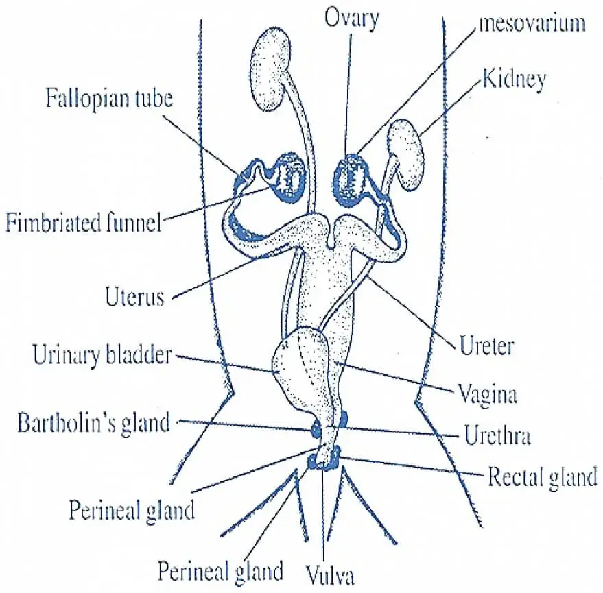
The female reproductive organs include a pair of ovaries, a pair of oviducts, a pair of uteri, a vagina, a vestibule, a clitoris, and some accessory glands.
Ovaries
- The two ovaries are tiny, whitish, oval bodies, about 2 cm long.
- They are found behind the kidneys, each ovary attached to the dorsal abdominal wall by a double fold of peritoneum called mesovarium.
- From the surface of ovaries project several blister-like, small, rounded, semitransparent projections, called ovarian or Graafian follicles, each containing a developing ovum.
- Histologically, the section of a rabbit ovary shows a peripheral layer of germinal epithelial cells surrounding a dense mass of connective tissue fibers, called stroma, containing blood lymph vessels, and nerves.
- Stroma contains groups of actively dividing germinal cells, called follicles in various stages of development.
- In each follicle, a single cell enlarges first while others surround and nourish it. It ultimately becomes an oocyte or ovum.
- The mass of cells around the oocyte is known as discus proliferous.
- When ripe, the follicles are known as Graafian follicles, which project from the surface of the ovary as minute bumps.
- Each Graafian follicle contains a large fluid-filled follicular cavity.
- The cells lining the cavity are termed membrane granulosa.
- The fully mature oocyte is surrounded by a thick transparent membrane called zona pellucida containing yolk and fat droplets.
- It is covered by another striated layer of columnar cells, called corona radiata.
- In the stroma, there are also found groups of interstitial cells that produce sex hormones (estrogen).
- Eventually, each mature follicle bursts to liberate the oocyte into a body cavity, a process known as ovulation.
- The follicular cells remaining behind divide rapidly to form a yellowish solid mass of cells called corpus luteum.
- During pregnancy, it serves as a temporary endocrine gland secreting a hormone (progesterone).
- It causes the uterus to enlarge to receive the growing fetus and stimulates lactation.
- If the ovum is not fertilized, the corpus luteum gradually disappears leaving a scar called corpus albicans.
Oviducts
- Each oviduct opens anteriorly, close to the outer border of the ovary of its side, by a wide funnel called fallopian or oviducal funnel.
- The opening of the funnel, or ostium, is provided with many cilia to receive the minute ova released from the ovary.
- The funnel leads into the upper part of the oviduct. It is a short, narrow, coiled, and internally ciliated duct called a fallopian tube. Ova passes through this tube by ciliary action and fertilization also occurs here.
- The fallopian tube is followed by a much broader, longer convoluted, thick-walled muscular tube the uterus.
- It is richly vascular and highly distensible and attached to the dorsal abdominal wall by a mesentery.
- Fertilized ova or zygotes get implanted on the uterine wall to develop into embryos or fetuses, each attached to the placenta by an umbilical cord.
Vagina and Vestibula
- The uteri of both sides meet into a long wide, median duct, the vagina, lying dorsally upon the urinary bladder.
- It opens posteriorly into the neck of the bladder to join the urethra forming a short narrow standard urinogenital canal or vestibule.
- It runs backward ventral to the rectum and opens to the exterior by a slit-like aperture, the vulva.
- The vagina serves to receive the penis of the male during copulation.
Clitoris
- The anterior wall of the vulva projects a small erectile knob-like clitoris.
- It is regarded as homologous with the male penis since it contains a pair of erectile tissue, the corpora cavernosa.
- However, the urethra does not pass through the clitoris.
Accessory Glands
- In the female rabbit, there is no prostate gland.
- A pair of small Bartholin’s glands or Cowper’s glands lies embedded in the dorsal wall of the vestibule.
- Their viscid secretion lubricates the vaginal passage. The perineal and rectal glands are as in the male.
Medically Reviewed
The information on this page is peer reviewed by a qualified editorial review board member. Learn more about us and our editorial process.
Last reviewed on .
Article history
- Latest version
Cite this page:
- Comment
- Posted by Dayyal Dungrela
Start a Conversation
Add comment
Follow us on social media
End of the article