Contraction of Muscle
Explore the mesmerizing world of muscle contraction, from actin and myosin to ATP and nervous control. Uncover nature's precision in motion.
Muscle contraction is an intricate biological process that lies at the heart of our ability to move, breathe, and perform countless other essential functions. This remarkable phenomenon involves a highly coordinated interplay of molecular and cellular events, and its understanding has been a subject of fascination for biologists and researchers for centuries.
The Molecular Ballet: Actin and Myosin
At the core of muscle contraction are two key proteins: actin and myosin. These proteins act as the primary players in the molecular ballet that powers muscle movement. Actin forms thin filaments, while myosin forms thick filaments within muscle fibers. The interaction between these filaments is akin to a finely choreographed dance.
When a signal from the nervous system instructs a muscle to contract, it triggers a release of calcium ions within the muscle cell. These ions bind to regulatory proteins, allowing actin and myosin to interact. Myosin has tiny "arms" that reach out and attach to actin, forming cross-bridges. In a process fueled by adenosine triphosphate (ATP), myosin pulls on actin, causing the filaments to slide past each other. This action shortens the muscle, resulting in contraction.
Sliding Filaments: The Sarcomere
The fundamental unit of muscle contraction is the sarcomere. It's the segment of muscle fiber where the sliding filament theory unfolds. Within the sarcomere, actin and myosin filaments overlap, creating a striated pattern that's characteristic of muscle tissue. As myosin pulls on actin, the sarcomere shortens, and this contraction propagates along the entire muscle fiber.
The sliding filament theory beautifully illustrates the precision of nature's design. Muscle contraction occurs with astonishing speed and accuracy, allowing us to perform delicate tasks like threading a needle or executing powerful movements such as lifting weights.
Energy and Control: ATP and Nervous System
Muscle contraction demands a significant amount of energy, primarily supplied by ATP. This molecule is the energy currency of cells and is vital for myosin to continue its "pulling" action. The nervous system plays a crucial role in regulating muscle contractions. Motor neurons transmit signals from the brain to muscle fibers, determining the strength and duration of contractions.
The Complexity of Muscle Contraction
While this article provides a simplified overview, the reality of muscle contraction is far more complex. Different muscle types (skeletal, smooth, and cardiac) exhibit unique contraction mechanisms. Factors like muscle fatigue, oxygen availability, and muscle fiber types also influence the process.
The contraction of muscle is a captivating biological phenomenon that underscores the elegance of nature's design. The intricate interplay of proteins, the precision of the sarcomere, the role of ATP, and the control of the nervous system together orchestrate our every movement. Studying muscle contraction not only enhances our understanding of physiology but also inspires awe for the wonders of the human body.
- Contraction is the main property of muscle.
- The muscle receives stimuli through the motor nerve.
- The teledendrites of the last neurons of the motor nerve, which innervate in the muscle are called "neuromotor end plates".
- When the nerve impulse passes to the neuromotor end plates, "acetylcholine" is released.
- Acetylcholine stimulates the muscle to contract.
- After the muscle starts contraction acetyl Choline is destroyed immediately by "choline esterase".
- The muscle contracts totally or does not contract.
- The strength of contraction depends upon the intensity of the stimulus.
- Muscle contracts due to nerve impulses.
- But for stimulation to contract, the muscle fiber always requires a specific minimum strength of nerve impulse or stimulus.
- This is called "Threshold stimulus".
- The single isolated contraction of muscle fiber is called "Muscle twitch'.
- When the muscle fiber is stimulated by rapid nerve impulses, the muscle fiber remains in a state of contraction so long as the stimulation is continuous.
- Such a continued state of contraction is called "Tetanus".
- The muscle fiber always contracts with maximum force irrespective of the intensity of the stimulus.
- The force of contraction does not increase even though the intensity of the stimulus is increased.
- This is known as the "all-or-none law". But muscle does not contract when the stimulus is below the threshold stimulus.
- The muscles which contract to produce opposite movements at the same joint are called "Antagonistic muscles".
- When a muscle contracts, its antagonistic muscle must relax to allow the movement.
- The biceps is a flexor muscle that, folds the elbow joint and the triceps is an extensor, the muscle that extends the elbow joint.
- Related Resources: Structure of the muscle
Mechanism of Muscle Contraction
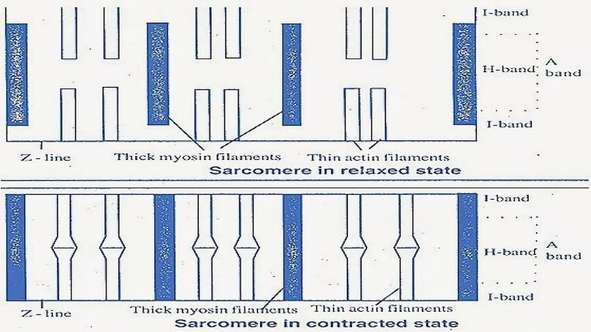
- When the muscle is in a relaxed state, I- bands and H - bands are longer, and A - bands or thick bands are shorter, When the muscle is in a contracted state, I - bands and H - bands are smaller but A -bands or thick bands are larger.
- This change occurs in the entire length of the sarcomere.
- The muscle contraction was explained by sliding filament theory.
Sliding Filament Theory
- It was proposed by H.E. Huxley and A.F. Huxley [Jean Hanson].
- According to them, the muscle becomes shorter and shorter, the thick and thin filaments slide past each other.
- However, the length of the filaments is not altered. As the thin filaments slide past the thick filaments, the thin filaments] meet in the center of the sarcomere.
- Therefore the TV band never disappears.
- But the l-band shortens and disappears.
- The actual sites of contraction are the cross-bridges, which are formed when the two filaments overlap. During relaxation, the cross bridges break.
- Hence the thin actin filaments move away from the 'A' band. Then the l-band reappears.
- Each thick or myosin filament is surrounded by six actin filaments in hexagonal form.
- Each cross-bridge is attached to an actin filament.
- At this place of contact, actin, and myosin filaments are attached to each other forming actomyosin which initiates contraction, i.e. the actin filaments slide past the thick myosin filaments.
- Each cross-bridge is formed of one ATP molecule. For every second cross bridge disappears and appears 50 to 100 times
Ratchet Meatchet Mechanism
- During contraction and relaxation, the cross bridges operate in a cyclic manner alternately attaching and detaching from thin actin filaments.
- This is called the ratchet mechanism. The bridges act like hooks or levers.
- A number of mitochondria are present in between the filaments which provide ATP necessary for muscle contraction.


Chemical Events
During muscle contraction, there occurs many chemical events. These are first worked out by "Szent Gyorgi."
- Whenever the acetylcholine is released on the muscle, the Ca++ is released from the endoplasmic reticulum [sarcoplasmic reticulum]. Acetylcholine + E.R → Ca++
- The enzyme myosin A.T.P. ase is active in the presence of Ca++, and Mg++. Which are also essential for muscle contraction. The A.T.P. ase breaks ATP into ADP, P [phosphate], and energy in the presence of Ca++ and Mg++ by hydrolysis. ATP + H2O → ADP + P + Energy
- The thick myosin filaments now bind the thin actin filaments by using energy. Hence "Actomyosin" is formed. Actin + Myosin → Actomyosin
- For relaxation, muscle requires energy. Hence ADP is immediately changed to ATP by creatine phosphate. ADP + CP → ATP + C [creatine]
- Now again the ATP breaks into ADP to release energy. Such energy is used to break down the actomyosin. Hence thin filaments move to the normal position. Thus the l-band reappears. Actomyosin + Energy → Actin + Myosin
- The creatine produced is converted into creatine phosphate by ATP. Creatine + ATP → ADP + CP
[creatine phosphate] - The ATP is synthesized by the oxidation of carbohydrates [glucose]. C6H12O6 + O2 → CO2 + H2O + ATP
- The muscle stores glucose in the form of glycogen. During strenuous exercise, the muscle does not get sufficient oxygen. Hence muscle respires by anaerobic glycolysis. Glycolysis in the muscle was explained by "Cor/and Corf. They proposed the glycolysis in muscle and liver as Cori's cycle. The muscle glycogen is broken down into lactic acid and energy. The energy is used for the synthesis of CP. The lactic acid is released into the blood, which is circulated to the liver. In the liver 4/5 of lactic acid is converted into glucose and 1/5 is oxidised to CO2 and H2O. The glucose is transported to the muscle through the blood. In muscle, glucose is converted into glycogen. The steps in Cori's cycle are given below.
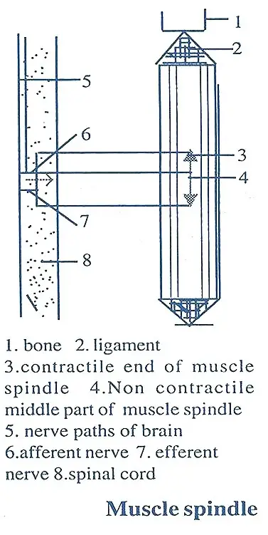
Stretch Reflex
- The muscle spindle is present parallel to the muscle fibers of a muscle.
- In the middle part of the muscle spindle, a bundle of small muscle fibers is present.
- They are non-contractile.
- The two ends of the muscle spindle contain spinal nerve endings and are contractile.
- The two contractile ends of the muscle spindle are supplied with afferent and efferent nerves from the central nervous system.
- As and when muscles expand, the nerve impulses are transmitted from muscle endings to the central nervous system through afferent nerves.
- The muscles receive responses accordingly from the CNS through efferent nerves.
- As a result, the two endings of the muscle spindle undergo contraction.
- This is called "stretch reflex".
The information on this page is peer reviewed by a qualified editorial review board member. Learn more about us and our editorial process.
Last reviewed on .
Article history
- Latest version
Cite this page:
- Comment
- Posted by Dayyal Dungrela