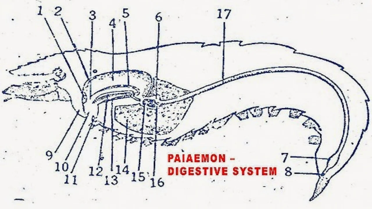
Digestive System of Palaemon (Prawn)
The digestive system of Palaemon (prawn) consists of a simple tube-like structure with specialized regions for ingestion, digestion, and absorption of nutrients, allowing for efficient processing of food.

Palaemon's digestive system contains a long alimentary canal and a large hepatopancreatic gland.
the digestive system of Palaemon exhibits a well-organized and specialized structure for efficient digestion and absorption. From the mouth to the hindgut, each organ and region plays a vital role in breaking down food, extracting nutrients, and eliminating waste. Understanding the intricacies of the prawn's digestive system provides insights into its feeding habits and adaptations to its aquatic lifestyle.
I. Alimentary Canal
- The alimentary canal is a long tube.
- It starts at the mouth and ends with the anus.
- It shows the buccal cavity, esophagus, stomach intestine, and rectum.
- The buccal cavity, esophagus, and stomach are lined with euncle.
- It is called stomodaeum or fore-gut. The intestine is lined by endoderm and is called mesenteron or mid-gut.
- The rectum is lined by a cuticle and is called the proctodaeum or hindgut.
Mouth
- The mouth is a longitudinal slit on the ventral side of the head.
- It shows a labrum on the anterior side.
- Mandibles are lateral.
- The thin labium is present on the posterior side.
Buccal Cavity
- The mouth leads into the buccal cavity.
- It is short and vertical. It shows a thick and folded cuticle.
- The molar processes of the mandibles project into the buccal cavity.
- The buccal cavity opens into the esophagus.
Oesophagus
- It is a short and wide tube.
- Its wall is folded. There are 4 longitudinal folds.
- Each lateral fold is subdivided by a groove into two smaller olds.
- The cuticular lining of the esophagus bears bristles.
- The esophagus leads into the stomach.
Stomach
- It is a large sac.
- It occupies more than half of the cephalothorax region.
- It is divided into large cardiac and small pyloric regions.
Cardiac Stomach
- It is lined with a thin cuticle, it is longitudinally folded. In places, the cuticle is thickened and calcified into plates.
- Near esophageal opening circular plate is present.
- On the roof near the anterior end, a lanceolate plate is present.
- On the floor of the cardiac stomach, the hastate plate is present.
- Below the hastate plate, a pair of comb plates are present. The lateral groove separates the hastate plate and comb plate.
- Each comb plate has a dense fringe of delicate bristles, which are directed inwards.
- Each lateral groove has a groove plate on the floor.
- Lateral longitudinal folds or guiding ridges are present on either side of the comb plates.
- The cardio pyloric aperture is X-shaped and is bounded by anterior, posterior, and lateral valves.
- The margins of the valves bear setae which act as a sieve. It permits fluid or very fine food particles to pass into the pyloric stomach.
Pyloric Stomach
- It lies beneath the posterior part of the cardiac stomach.
- Its lateral walls are thick and project as large longitudinal folds into its lumen. They divide the pyloric stomach into a small dorsal chamber and a large ventral chamber.
- The lateral grooves on the sides of the hastate plate open into the ventral pyloric chamber.
- On the floor of the ventral chamber, thick plates are present side by side.
- They are "V" shaped In cross-section. These two plates are called filter plates. On the ridge of the filter plate, a row of bristles can be seen.
- The cuticle of the lateral walls of the ventral pyloric chamber and the filter plate forms a filter that permits only the fluid to pass through it.
- Behind the filter plate, pair of hepatopancreatic ducts will open into the ventral pyloric stomach.
Intestine
It is a long tube extending up to the 6th abdominal segment.
Rectum
It is very short. It extends from the 6th abdominal segment to the anus. Its anterior part is enlarged into a muscular sac.
Anus
It is present on the ventral side of the telson.
II. Hepatopancreas
- It is a large orange-red gland.
- It consists of two separate lobes and develops as a pair of outgrowths.
- The hepatic caeca. from the mid-gut.
- It lies around the stomach.
- It has many branching tubules held together by connective tissue.
- The tubules join to form larger tubes and they unite to form a pair of hepatopancreatic ducts.
- They open into the ventral pyloric chamber.
- It serves like the liver and pancreas of higher animals.
Functions
- It secrets digestive enzymes.
- It stores glycogen, fat, and calcium like the liver.
- It absorbs digested food from the intestine.
Physiology
Palaemon is omnivorous. It eats algae, weeds, insect larvae, small fish, and debris.
Ingestion
- Small food particles are caught by the chelate legs and pushed into the mouth.
- The 3rd maxillipeds assist the chelate legs in handling large food masses.
- The coxae of the 2nd, maxilliped will hold the food.
- The incisor processes of the mandibles cut the food into small bits.
Digestion
- Much of the digestion of food takes place in the cardiac stomach.
- It is brought by the digestive juice secreted by the hepatopancreas.
- Dissolved food passes into the lateral grooves and then into the ventral chamber of the pyloric stomach through the cardio-pyloric aperture.
- Digestion continues in the pyloric stomach.
- The food is filtered through the pyloric filter.
Absorption
- Absorption of food takes place in the hepatopancreas and intestine.
- The major part of the digested food passes into the hepatopancreas through the hepatopancreatic ducts.
- The undigested food particles left by the pyloric filter pass into the dorsal pyloric chamber.
- Then they go to the intestine.
Egestion
The undigested food is sent out through the anus.
The information on this page is peer reviewed by a qualified editorial review board member. Learn more about us and our editorial process.
Last reviewed on .
Article history
- Latest version
Cite this page:
- Comment
- Posted by Dayyal Dungrela