Pandy’s Test: Procedure, Interpretation & Clinical Significance
Pandy’s test detects elevated CSF proteins to diagnose CNS disorders. Learn how this biochemical assay works and its clinical applications.
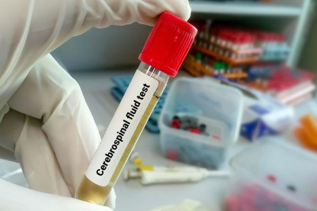
Pandy’s test (also called Pandy’s reaction) is a biochemical assay to detect elevated cerebrospinal fluid (CSF) proteins, particularly globulins. This test was originally developed by a Hungarian neurologist, Pándy Kálmán (1868–1945) in the yaer 1910. It is used to identify pathological conditions affecting the central nervous system (CNS).
What is the Pandy’s Test?
The Pandy’s test detects abnormal protein levels in CSF using phenol to precipitate globulins and albumin. A positive result indicates CNS disorders like meningitis or multiple sclerosis. While newer methods exist (e.g., electrophoresis), it’s still used in resource-limited settings.
Principle
The test relies on the precipitation of CSF proteins, globulins and albumin, when exposed to a saturated aqueous solution of phenol. Phenol disrupts the solubility of these proteins, causing them to aggregate and form visible turbidity or precipitates. Alternative reagents, such as pyrogallic acid or cresol, may also be used, though phenol-based solutions (termed Pandy’s reagent) are most common.
Procedure
- Sample Collection: CSF is obtained via lumbar puncture, a sterile procedure involving needle insertion into the subarachnoid space of the spinal canal.
- Reagent Preparation: Pandy’s reagent is prepared by dissolving carbolic acid crystals in water to create a saturated phenol solution.
- Testing: One drop of CSF is added to approximately 1 mL of Pandy’s reagent. The mixture is observed for turbidity development.
- Positive Result: Turbidity or a milky precipitate indicates elevated globulin levels.
- Negative Result: A clear solution reflects normal CSF protein content.
Interpretation of Results
Normal CSF Composition
In healthy adults, CSF contains proteins at a concentration of 15–45 mg/dL, with an albumin-to-globulin ratio of 8:1. Normal CSF remains clear and transparent.
Positive Pandy’s Reaction
A positive result manifests as a bluish-white streak or turbidity, with intensity correlating to protein concentration:
- Mild Turbidity: Suggests moderate protein elevation (e.g., 50–100 mg/dL).
- Dense Milky Precipitate: Indicates severe hyperproteinorrhea (>100 mg/dL).
Clinical Significance of Elevated CSF Proteins
A positive test may signal pathologies such as:
- Infectious Disorders: Acute purulent meningitis, granulomatous meningitis, or neurosyphilis.
- Neoplastic Conditions: Meningiomas, spinal cord tumors, or carcinomatous meningitis.
- Autoimmune/Inflammatory Diseases: Multiple sclerosis, Guillain-Barré syndrome, or connective tissue disorders.
- Metabolic/Endocrine Dysfunction: Diabetes mellitus, uremia, or myxedema.
- Vascular or Structural Abnormalities: Cerebral hemorrhage, brain abscesses, or subarachnoid block.
“For a comprehensive overview of CSF analysis in diagnosing these conditions, see our detailed guide on Examination of Cerebrospinal Fluid (CSF).”
Negative Pandy’s Reaction
Absence of turbidity confirms normal protein levels. However, certain pathologies such as viral CNS infections, ischemic strokes, or brainstem gliomas—may present with normal CSF protein, necessitating adjunct diagnostic tests.
Limitations and Considerations
While Pandy’s test is rapid and cost-effective, it is semi-quantitative and lacks specificity for individual protein subtypes. False negatives may occur in chronic syphilis or early-stage Guillain-Barré syndrome, where protein elevation lags symptom onset. Modern techniques like electrophoresis or immunoassays are often employed for precise quantification.
Takeway
- Use Pandy’s test for rapid CNS screening.
- Confirm positives with electrophoresis.
FAQs
What diseases does a positive Pandy’s test indicate?
How long does the Pandy’s test take?
Cite this page:
- Posted by Dayyal Dg.