Total Erythrocyte Count (Manual Method)
Learn detailed procedures for accurate red blood cell count estimation using microscopic techniques. Understand calculations and discover the causes of low red blood cell count and high red blood cell count.

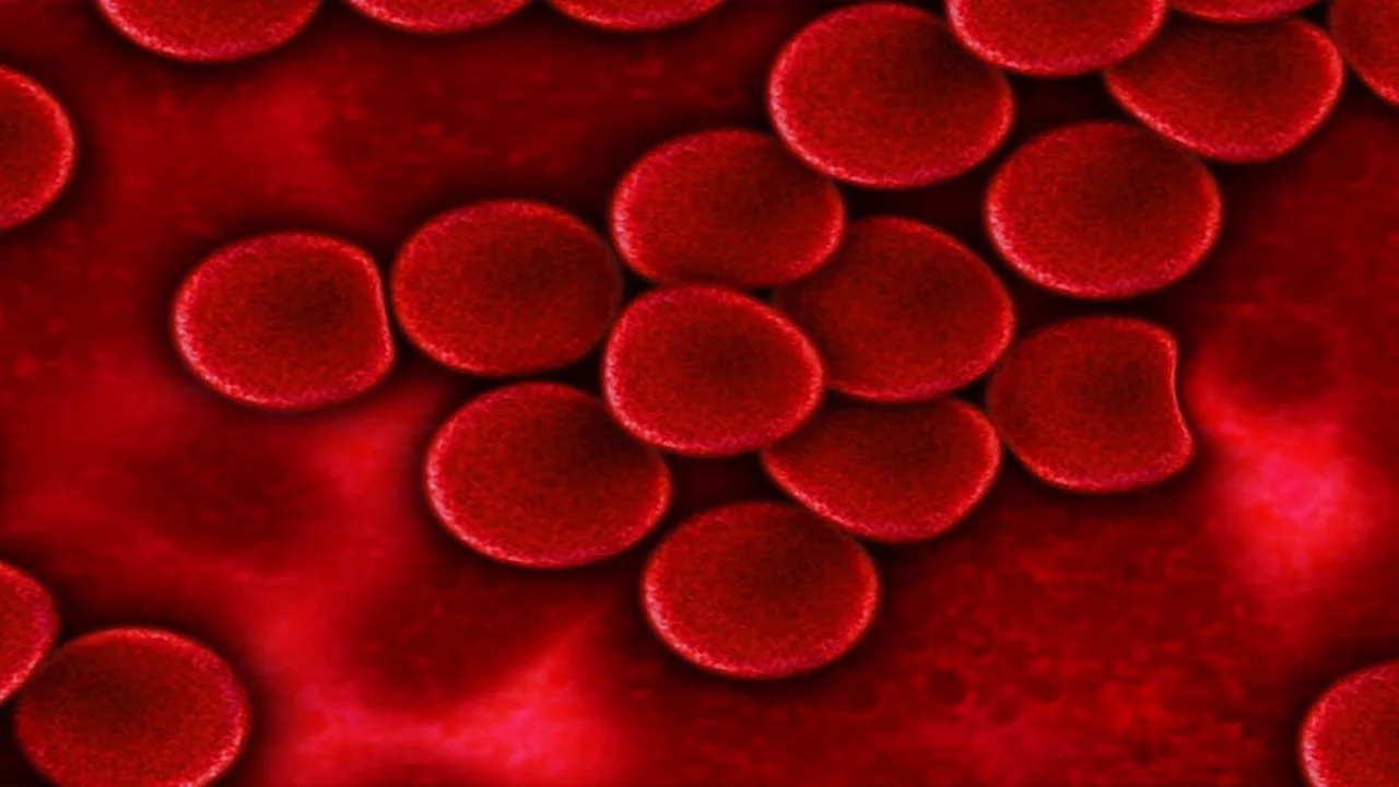
Erythrocytes (derived from Greek erythros meaning red, and kytos meaning cell), commonly known as red blood cells (RBCs), are circular, anucleated cells with a high degree of flexibility and a distinct biconcave disc shape. These cells measure approximately 7.2 micrometers in diameter and 2.1 micrometers in thickness, with a central thickness of about 1.0 micrometer. Encased in a complex bimolecular membrane of proteins, erythrocytes primarily contain a stroma made of lipids and fibrous proteins, with about 90% of the cell's content being hemoglobin. The erythrocyte count can be estimated using two primary methods:
- Manual (Microscopic) Method
- Automated Method
Red Blood Cell Count by Manual Method
Requirement for Red Blood Cell Count by Microscopic Method
Equipment
- Hemocytometer with cover glass
- Compound microscope
Reagent
RBC Diluting Fluid (Hayem’s Solution) is used for diluting RBC's. The Hayem’s solution contains mercuric chloride (HgCl₂), sodium sulfate (Na₂SO₄) and sodium chloride (NaCl). Chemical composition of Hymem’s diluting solution (RBC diluting fluid):
- Mercuric chloride (HgCl₂): 0.05 g
- Sodium sulfate (Na₂SO₄): 2.5 g
- Sodium chloride (NaCl): 0.5 g
- Distilled water: 100 ml
Specimen
EDTA-anticoagulated venous blood or capillary blood obtained via skin puncture.
Procedure
- Preparation:
- Clean the fingertip with alcohol-soaked cotton and make a small puncture with a sterile lancet. Use a pipette to aspirate blood to the 0.5 mark.
- Aspirate Hayem’s solution up to the 101 mark to achieve a 1:200 blood dilution.
- Mixing: Hold the pipette horizontally and roll it between the fingers and thumb to mix.
- Loading the Chamber:
- Place a dust-free, grease-free counting chamber on a table and cover it with a cover glass.
- Discard the first few drops from the pipette, then charge the counting chamber by holding the pipette at an inclined position and allowing 3 minutes for the cells to settle.
- Counting:
- Locate the central square, subdivided into 25 medium squares, each further divided into 16 smaller squares.
- Using a high-power objective, count the erythrocytes in the medium squares (80 smallest squares).
- Count cells touching the upper and left outer lines of each square but exclude those touching the lower and right inner lines.

Calculation
- Count the erythrocytes in five medium squares (E).
- Area of each square: 1/25 sq mm.
- Total area counted: 1/25 sq mm × 5 = 1/5 sq mm.
- Depth of the chamber: 1/10 mm.
- Volume counted: Area × Depth = 1/5 sq mm × 1/10 mm = 1/50 cu mm.
- Number of erythrocytes in 1/50 cu mm of diluted blood: E.
- Number of erythrocytes in 1 cu mm of diluted blood: E × 50
- Undiluted blood erythrocyte count: E × 50 × 200.
Polycythemia (High Red Blood Cell Count) Causes
- Congenital heart disease
- Cor pulmonale
- Dehydration
- Pulmonary fibrosis
- Polycythemia vera
Anemia (Low Red Blood Cell Count) Causes
- Anemia
- Bone marrow failure
- Erythropoietin deficiency (secondary to kidney disease)
- Hemolysis due to transfusion reactions
- Hemorrhage
- Leukemia
- Multiple myeloma
- Nutritional deficiencies (Iron, Copper, Folate, Vitamins B₁₂ and B₆)
Reference Ranges
- Newborns: 4.8-7.2 million cells/cu mm
- Children: 3.8-5.5 million cells/cu mm
- Adult Males: 4.6-6.0 million cells/cu mm
- Adult Females: 4.2-5.0 million cells/cu mm
- Pregnancy: Slightly lower than normal levels
The information on this page is peer reviewed by a qualified editorial review board member. Learn more about us and our editorial process.
Last reviewed on .
Article history
- Latest version
Reference(s)
- Brown, B.A., Haemotology, Principles and Procedures, Lea & Febiger, U.S.A., 1976.
- Hoffbrand, A. V. and Pettit, 1. E., Essential Haemotology, Blackwell Scientific Publication, U.S.A., 1980.
- Kassirsky, I. and Alexeev, G., Clinical Haemotology, Mir Publishers, U.S.S.R., 1972.
- Widmann, F.K., Clinical interpretation of Laboratory tests, F.A. Davis Company, U.S.A., 1985.
- Kirk, C.J.C. et al, Basic Medical Laboratory Technology, Pitman Book Ltd., U.K. 1982.
- Green, J.H., An Introduction to human Physiology, Oxford University Press, U.K., 1980.
Cite this page:
- Comment
- Posted by Dayyal Dungrela