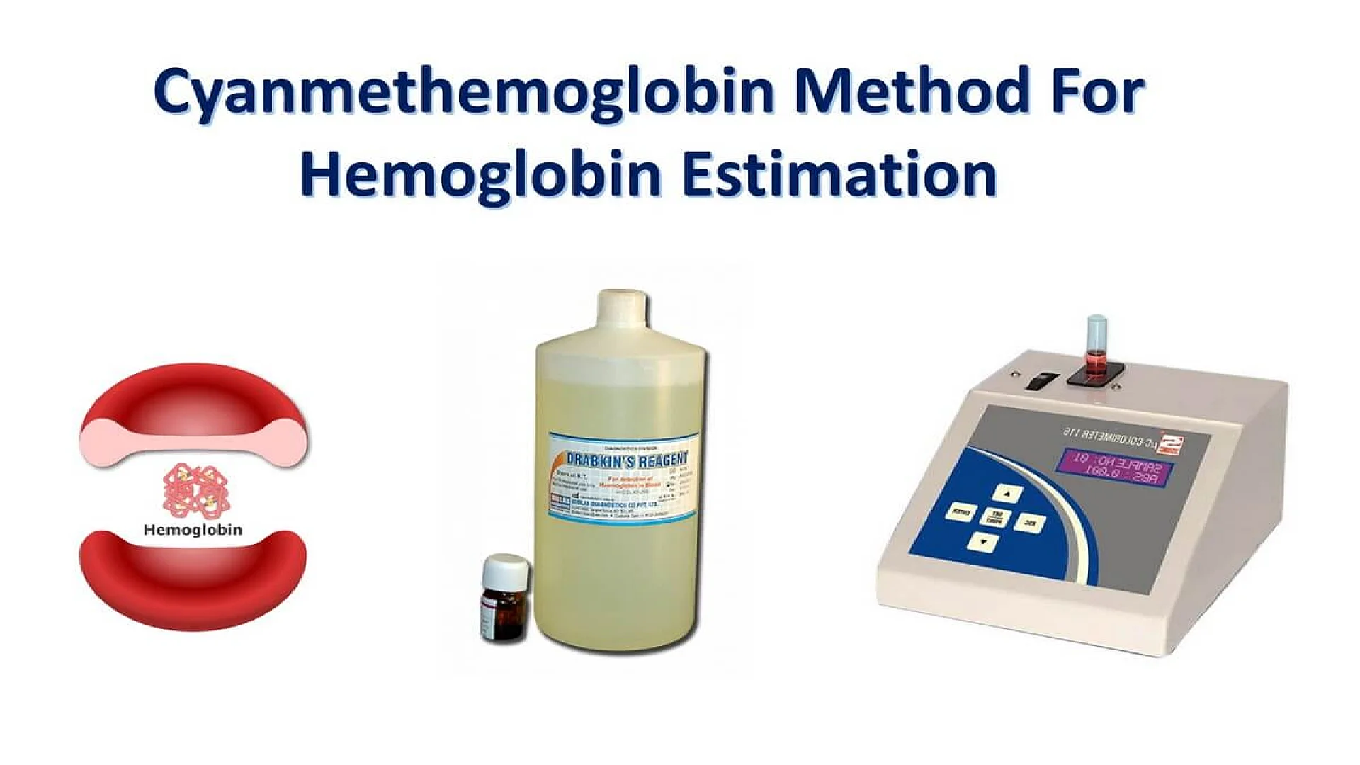
Cyanmethemoglobin (Hemoglobin Cyanide) Method for Estimation of Hemoglobin
Cyanmethemoglobin method for precise hemoglobin measurement. Includes procedure, formula, and reagents. Discover how to perform this standardized lab test.
Highlights
- What You’ll Read: Cyanmethemoglobin method principle using Drabkin’s reagent for precise hemoglobin estimation via spectrophotometry.
- What You’ll Learn: Procedure involves cyanmethemoglobin standard solution, reagent steps, and absorbance-based calculation formula.
- What You’ll Discover: ICSH standardization ensures global reproducibility despite leukocytosis or lipemia affecting accuracy.
The cyanmethemoglobin method is the gold-standard laboratory technique for estimating hemoglobin concentration in blood. Hemoglobin (Hb), a protein in red blood cells, carries oxygen and maintains blood pH. This method converts hemoglobin into a stable compound for accurate spectrophotometric analysis, recommended by global hematology standards.
Hemoglobin is made up of four heme molecules, each attached to a globin chain. In adults, three types of hemoglobin are present:
- HbA (96-98%): Composed of two alpha (α) and two beta (β) globin chains.
- HbA2 (1.5-3.5%): Composed of two alpha (α) and two delta (δ) globin chains.
- HbF (<1%): Composed of two alpha (α) and two gamma (γ) globin chains.
Hemoglobin can bind with other substances, both normally and abnormally, forming different compounds:
- Oxyhemoglobin: Hemoglobin bound to oxygen.
- Carboxyhemoglobin: Hemoglobin bound to carbon monoxide (CO).
- Carbaminohemoglobin: Hemoglobin bound to carbon dioxide (CO2).
- Methemoglobin: Hemoglobin with oxidized iron (from ferrous to ferric state).
- Sulfhemoglobin: Hemoglobin bound to sulfur.
- Cyanmethemoglobin: Hemoglobin bound to cyanide ions.
What is cyanmethemoglobin method?
The cyanmethemoglobin method measures hemoglobin by converting it into cyanmethemoglobin using potassium cyanide and ferricyanide in Drabkin’s solution, which lyses red blood cells. Absorbance is measured at 540 nm for precise quantification, as endorsed by the International Committee for Standardization in Hematology (ICSH).
History of Cyanmethemoglobin Method for Hemoglobin Estimation
The cyanmethemoglobin method for hemoglobin estimation was developed by Dr. Leonard Drabkin in the mid-20th century. Drabkin, an American hematologist and biochemist, introduced this method as a more accurate and reliable alternative to existing hemoglobin measurement techniques. His work with potassium cyanide and potassium ferricyanide solutions, which lysed red blood cells to create a stable cyanmethemoglobin complex, became foundational in hematology.
This method was later adopted by the International Committee for Standardization in Hematology (ICSH) as the preferred standard due to its precision and effectiveness in transforming all major forms of hemoglobin (except sulfhemoglobin) into a single, measurable compound. Drabkin’s contribution revolutionized clinical diagnostics by providing a dependable technique for hemoglobin estimation used worldwide today.
Advantages of Cyanmethemoglobin Method for Hemoglobin Estimation
The cyanmethemoglobin method is the preferred technique for hemoglobin estimation due to its significant advantages, including accuracy, standardization, and cost-effectiveness:
- Accuracy and Precision: Converts most hemoglobin types into a single stable compound for consistent results.
- Broad Detection: Measures all hemoglobin forms except sulfhemoglobin.
- Standardization: Globally recognized as the reference method by ICSH.
- Ease of Use: Compatible with automated spectrophotometric analyzers.
- Stability: Cyanmethemoglobin remains stable for hours, reducing processing errors.
- Cost-Effectiveness: Uses affordable reagents like Drabkin’s solution, ideal for routine testing
Principle of Cyanmethemoglobin Method
The principle of the cyanmethemoglobin method involves converting all hemoglobin types in a blood sample into cyanmethemoglobin, a stable compound, using potassium cyanide and potassium ferricyanide in Drabkin’s solution. The absorbance of cyanmethemoglobin is measured at 540 nm using a spectrophotometer, and the hemoglobin concentration is determined by comparing this absorbance to a standardized solution. This method ensures precise, consistent, and reliable hemoglobin estimation.
Step 1: Conversion of Hemoglobin to Methemoglobin
Equation: Hb + K₃Fe(CN)₆ → MetHb + K₄Fe(CN)₆
In this reaction, potassium ferricyanide (K₃Fe(CN)₆) oxidizes hemoglobin (Hb) to methemoglobin (MetHb).
Step 2: Formation of Cyanmethemoglobin (Hemiglobincyanide)
Equation: MetHb + KCN → Cyanmethemoglobin (Hemiglobincyanide)
Here, methemoglobin (MetHb) reacts with potassium cyanide (KCN) to form cyanmethemoglobin, the stable colored compound measured in the assay.
Required Equipment
- Spectrophotometer or photoelectric colorimeter.
- Pipette 5 ml.
- Sahli’s pipette.
Reagents
- Drabkin’s Solution (pH 7.0-7.4):
- Potassium ferricyanide 200 mg
- Potassium cyanide 50 mg
- Potassium dihydrogen phosphate140 mg
- Non-ionic detergent 1 ml
- Distilled water 1000 ml
- Cyanmethemoglobin standard solution with a verified hemoglobin value.
Specimen
Blood samples may be obtained via skin puncture or from EDTA-anticoagulated venous blood.
Cyanmethemoglobin Method Procedure for Hemoglobin Estimation
Step-by-Step Procedure:
- Prepare the Blood Sample: Mix 5 ml of Drabkin’s solution with 20 μl of blood to create a 1:25 dilution. Stir the mixture and let it sit for at least 5 minutes to fully convert hemoglobin to cyanmethemoglobin.
- Measure Absorbance: Transfer the mixture to a cuvette and measure absorbance at 540 nm using a spectrophotometer. Alternatively, use a photoelectric colorimeter with a yellow-green filter. Ensure the measurement is taken against Drabkin’s solution as a reference.
- Calculate Hemoglobin Concentration: Use the following formula to calculate hemoglobin concentration:
| Component | Blank | Standard | Test |
|---|---|---|---|
| Drabkin’s Reagent | 5 ml | 5 ml | 5 ml |
| Hemoglobin Standard | – | 20 µl | – |
| Sample | – | – | 20 µl |
Calibration Table and Graph Preparation
For efficient hemoglobin quantification, a calibration graph is essential, especially in laboratories processing high sample volumes. Here’s how to prepare the graph:
- Prepare Standard Solutions: Use commercially available adulterated cyanmethemoglobin standards or create a serial dilution of standard cyanmethemoglobin in Drabkin’s solution.
- Plot the Graph: On linear graph paper, plot the hemoglobin concentration (horizontal axis) against the corresponding absorbance (vertical axis). The points should form a straight line passing through the origin.
- Generate the Calibration Table: This graph can be used to create a calibration table, which correlates absorbance values with hemoglobin concentrations, aiding in rapid and accurate data interpretation.
Factors Affecting Cyanmethemoglobin Method Accuracy
- The cyanmethemoglobin solution remains stable, allowing flexible timing for absorbance readings without affecting accuracy.
- High white blood cell counts (TLC > 25,000/μl), abnormal plasma proteins (e.g., in Waldenström’s macroglobulinemia or multiple myeloma), and lipemic blood (hypertriglyceridemia) can cause errors in hemoglobin measurements.
FAQs
What is the cyanmethemoglobin method?
How does the cyanmethemoglobin method work?
Who developed the cyanmethemoglobin method?
What precautions should be taken when using the cyanmethemoglobin method?
What is hemiglobincyanide?
The information on this page is peer reviewed by a qualified editorial review board member. Learn more about us and our editorial process.
Last reviewed on .
Article history
- Next review due:
- Latest version
Reference(s)
- Cheesbrough, Monica. “District Laboratory Practice in Tropical Countries.” 2nd ed., Cambridge University Press, 2006, isbn: 9780511543470. <https://doi.org/10.1017/CBO9780511581304>.
- SM, Lewis., et al. “Dacie and Lewis’s Practical Haematology.” 9th ed., Churchill Livingstone, 25 Oct. 2001, isbn: 9780443063770. <https://shop.elsevier.com/books/dacie-and-lewis-practical-haematology/bain/978-0-7020-6696-2>.
Cite this page:
- Posted by Dayyal Dungrela
- Cell Biology
- Cyanmethemoglobin Apparatus
- Cyanmethemoglobin Method
- Cyanmethemoglobin Method Hemoglobin Estimation
- Cyanmethemoglobin Method Procedure
- Cyanmethemoglobin Method Sources of Error
- Cyanmethemoglobin MSDS
- Cyanmethemoglobin Reagent
- Cyanmethemoglobin Standard
- Cyanmethemoglobin Standard Solution
- Hemotological Tests
- Hemotology
- How-to
- Microbiology
- Zoology