Micro Method for Estimation of Hematocrit (HCT) or Packed Cell Volume (PCV)
Learn the principles, equipment, specimen handling, and detailed method for accurate hematocrit determination. Understand the reference ranges and critical values for optimal clinical interpretation.
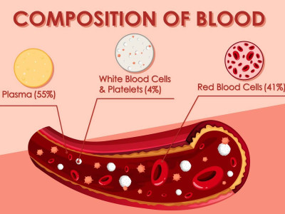
Hematocrit, expressed as a percentage, indicates the volume of red blood cells in relation to the total blood volume, which includes red blood cells, white blood cells, and platelets suspended in plasma. Typically constituting around 45% of blood volume, the precise distribution of these components can vary. This measurement, derived from a straightforward blood test, plays a crucial role in assessing the oxygen-carrying capacity of red blood cells throughout the body. Deviations from normal levels in hematocrit readings may indicate potential blood disorders or other medical conditions.
Principle of Hematocrit Determination
The process begins with centrifuging anticoagulated whole blood in a capillary tube of uniform bore to densely pack the red cells. This centrifugation occurs in a specialized microhematocrit centrifuge until the red cells are packed as completely as feasible. Measurements of the packed red cell length and the total column length are obtained using a microhematocrit reader, a ruler, or arithmetic graph paper.
Equipment Required for Hematocrit Testing
- Microhematocrit centrifuge: This equipment should provide a relative centrifugal force of 12,000 g for a duration of 5 minutes.
- Capillary hematocrit tubes: These disposable glass tubes measure 75 mm in length and have a 1 mm internal diameter. They are available in two types: plain (without anticoagulant) and heparinised (coated with a dried film of 2 units of heparin). Plain tubes require anticoagulated venous blood, while heparinised tubes are used for blood obtained from skin puncture.
- Tube sealant: Options include plastic sealant or modeling clay; alternatively, a spirit lamp can be used for heat sealing.
- Microhematocrit reader: If unavailable, measurements can be taken using a ruler or arithmetic graph paper.
Specimen Collection for Hematocrit Measurement
Venous blood is collected in EDTA (dipotassium salt) for plain tubes or directly into heparinised tubes from skin puncture. Collection should minimize stasis to prevent hemoconcentration and false elevation of packed cell volume (PCV).
Methodology for Hematocrit Determination
- Filling the capillary tube: Place the capillary tube tip in contact with the blood (either from skin puncture or anticoagulated venous blood, depending on the tube type). Fill approximately two-thirds to three-quarters of the tube length with blood.
- Sealing the tube: Seal the end of the capillary tube not in contact with blood using plastic sealant. Alternatively, heat seal the tube using a spirit lamp if plastic sealant is unavailable.
- Centrifugation: Position the filled tubes in the radial grooves of the centrifuge with sealed ends facing outward. Counterbalance by placing tubes in opposite grooves.
- Centrifugation parameters: Centrifuge at a relative centrifugal force of 12,000 g for 5 minutes to achieve maximal red cell packing.
- Reading the results: Immediately after centrifugation, stand the tubes upright. The tube contents will stratify into three layers: a plasma column at the top, a thin buffy coat layer, and a packed red cell column at the bottom.
- Hematocrit measurement: Use a microhematocrit reader to directly measure hematocrit from the scale. In the absence of a reader, hold the tube against a ruler and calculate hematocrit using the formula:
Additional Considerations and Interpretations
- Tourniquet application: Prolonged use during venepuncture may cause hemoconcentration, leading to an artificial rise in hematocrit.
- Finger squeezing: Excessive pressure during skin puncture can dilute the blood sample with tissue fluid, resulting in a lower hematocrit reading.
- Anticoagulant proportion: Proper mixing of blood with anticoagulant is crucial; excess EDTA can cause red cell shrinkage and falsely decrease hematocrit.
- Mixing errors: Inadequate mixing of blood before testing or with anticoagulant can lead to inaccurate hematocrit results.
- Clot interference: Presence of clots in the sample may lower hematocrit values.
- Centrifugation impact: Lower speed or insufficient centrifugation time can artificially increase PCV readings.
- Plasma trapping: Normally insignificant, but conditions like microcytosis, macrocytosis, spherocytosis, and sickle cell anemia can cause artifactual rises in hematocrit due to increased plasma trapping.
- Plasma volume effects: PCV is influenced by plasma volume changes, such as higher values in dehydration and lower in fluid overload.
- PCV expression: In SI units, PCV is expressed as a volume fraction (conversion factor: SI to conventional units is 100; conventional to SI units is 0.1).
- Rules of 3 and 9: These rules apply under specific conditions (normal red cell size and shape):
- Hemoglobin (g/dL) × 3 = PCV
- Red cell count (million/cmm) × 9 = PCV
- Automated hematocrit: Modern analyzers determine hematocrit by multiplying red cell count (in millions/cmm) by mean cell volume (in femtoliters).
Reference Ranges for Hematocrit
- Adult males: 40-50%
- Adult females (nonpregnant): 38 45%
- Adult females (pregnant): 36-42%
- Children 6 to 12 years: 37-46%
- Children 6 months to 6 years: 36 42%
- Infants 2 to 6 months: 32-42%
- Newborns: 44-60%
Critical Values for Hematocrit
- Packed cell volume: < 20% or > 60%
Cite this page:
- Comment
- Posted by Dayyal Dg.