Scientists Find This Bacterial Protein for Faster Intestinal Healing
A new study shows that intestinal stem cells can detect a bacterial protein and recruit helpful immune cells, strengthening the gut barrier and reducing inflammation during injury.
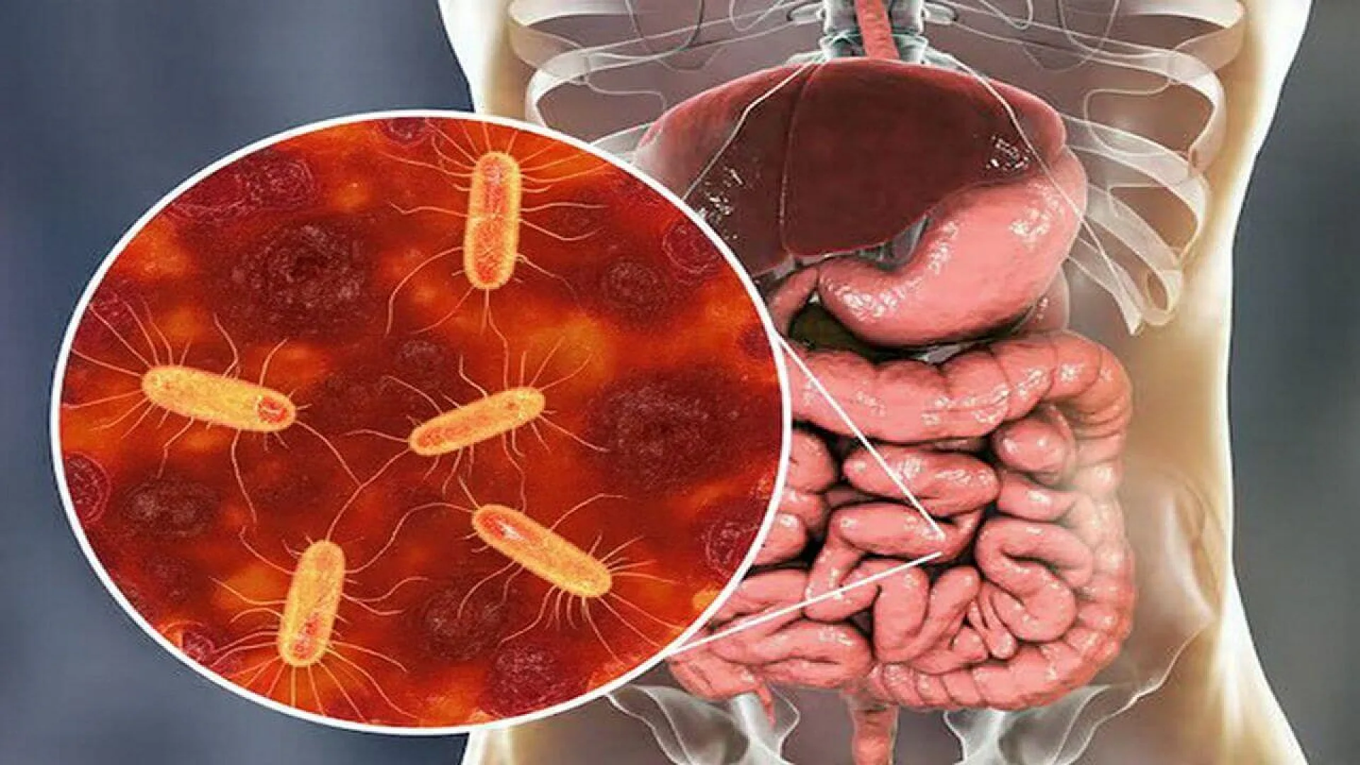
Inside the human intestine, a single layer of cells stands between the body and the outside world. On one side is the bloodstream and internal organs. On the other side are trillions of bacteria, viruses, and fungi that live in the gut.
This barrier is thin, and it is under stress all the time. Food passing through creates friction. Digestive chemicals can irritate cells. Bacterial products are always present.
Small injuries happen regularly. In healthy people, the intestine repairs itself quickly, often without any symptoms.
When this repair system breaks down, problems begin. Long-lasting damage to the gut lining is a key feature of inflammatory bowel diseases, such as ulcerative colitis and Crohn’s disease.
Just below the gut lining is a layer filled with immune cells. Among them are macrophages, large cells that clean up debris, remove microbes, and help tissues heal.
In a healthy intestine, these macrophages are calm and supportive. They do not cause unnecessary inflammation. Instead, they help maintain balance.
Macrophages in the gut are constantly replaced. New cells arrive from the blood as monocytes, which then mature inside the intestinal tissue.
Scientists already knew that gut bacteria are important for this process. Animals raised without microbes, or treated with strong antibiotics, have fewer of these helpful macrophages.
What was not clear was how specific bacteria trigger this immune support.
A Protective Strain of E. coli
The new study focused on a particular strain of Escherichia coli, known as 541-15. This bacterium belongs to a group that can closely attach to intestinal cells.
Earlier experiments had shown something unexpected. When mice carried this strain, they were more resistant to colitis, a condition marked by inflammation and damage in the colon.
The reason for this protection was unknown. The new research set out to find the mechanism.
Testing the Effects of One Bacterium
To begin, researchers treated mice with antibiotics. This reduced their normal gut microbes and also reduced protective macrophages in the intestine.
The scientists then introduced either E. coli 541-15 or a harmless control strain that does not protect against colitis.
Both bacteria were able to live in the gut. However, their effects were very different.
Only mice given E. coli 541-15 showed a recovery of mature intestinal macrophages.
Rebuilding the Immune Network
In these mice, macrophages formed a dense network just beneath the gut lining. This pattern is typical of a healthy intestine.
The control strain failed to restore this network, even though it was present in similar numbers.
Detailed cell analysis confirmed that E. coli 541-15 increased macrophages known to support tissue repair and reduce inflammation.
This showed that the effect was not due to bacteria in general. It depended on something specific to this strain.
Protection During Gut Injury
The researchers then tested whether these immune changes actually mattered.
They exposed the mice to a chemical called dextran sulfate sodium. This substance damages the gut lining and is commonly used to model colitis in animals.
Mice carrying E. coli 541-15 became less sick. They lost less weight, showed milder symptoms, and had less damage in their colon.
Their intestines were also longer, which is a sign of reduced inflammation. In addition, inflammatory markers in stool samples were lower.
Under the microscope, the gut tissue looked healthier, with more macrophages positioned close to the epithelium.
The Role of the Gut Lining Itself
Macrophages respond to signals from surrounding cells. In the intestine, many of those signals come from epithelial cells, the cells that form the gut lining.
To understand this interaction, researchers studied human colonic organoids. These are miniature gut-like structures grown in the lab from human tissue.
Some organoids were made mostly of mature epithelial cells. Others were enriched in undifferentiated cells, including intestinal stem cells found deep in gut crypts.
This difference turned out to be important.
Stem Cells Sense the Bacterium
When mature epithelial cells were exposed to E. coli 541-15, they showed little change in gene activity. But when undifferentiated cells were exposed to the same bacterium, their response was strong.
Hundreds of genes involved in immune signaling became active. One of the most important was CCL2, a chemical signal that attracts monocytes from the bloodstream.
These signals were released toward the tissue side of the epithelium, exactly where immune cells are located.
Calling Immune Cells to the Right Place
CCL2 acts like a guide. It helps monocytes find their way into the gut tissue, where they can mature into macrophages.
The study showed that this signal was essential. When mice lacked CCL2 specifically in their gut lining, E. coli 541-15 could no longer protect them.
Macrophages were not recruited properly, and gut damage increased. This confirmed that epithelial cells were actively directing the immune response.
A Key Protein Called Flagellin
Next, the researchers asked what part of the bacterium triggered this response.
They compared several E. coli strains and found a clear link. Strains that produced large amounts of flagellin caused strong immune signaling. Strains with little flagellin did not.
Flagellin is the protein that makes up the bacterial flagellum, a tail-like structure used for movement.
When the team used bacteria lacking flagellin, the protective effect disappeared. These bacteria could still live in the gut, but they no longer recruited macrophages or reduced colitis.
How the Gut Detects Flagellin
Intestinal cells detect flagellin using a receptor called Toll-like receptor 5, or TLR5. This receptor sits on the surface of epithelial cells and recognizes specific bacterial patterns.
When researchers removed TLR5 only from intestinal epithelial cells, E. coli 541-15 stopped working. Macrophage recruitment was reduced, and gut injury increased.
This showed that the gut lining itself must sense flagellin for the repair process to begin.
Why Stem Cells Matter So Much
The most responsive cells were intestinal stem cells and their early descendants. These cells live at the base of intestinal crypts and constantly produce new epithelial cells.
By sensing bacteria, these stem cells appear to coordinate repair from the very start of tissue renewal. They are not just passive builders. They help shape the immune environment needed for healing.
A Balanced Immune Response
Importantly, this pathway did not trigger harmful inflammation. Instead, it promoted a controlled immune response focused on repair.
This balance is essential. Too much inflammation can worsen damage. Too little leaves the gut vulnerable.
The findings help explain how certain microbes support health without causing disease.
What This Means for Gut Disease
The study helps explain why antibiotics can sometimes worsen intestinal problems. By disrupting specific microbes, they may break important repair signals.
It also suggests new treatment ideas. Instead of suppressing the immune system, future therapies might aim to restore helpful microbe–immune communication.
Questions Still to Answer
The research focused on one bacterial strain and controlled animal models. Human gut ecosystems are far more complex.
Scientists still need to learn whether similar mechanisms operate across different people and microbial communities.
It is also unclear how this pathway interacts with other bacterial signals in the gut.
A More Active View of the Gut
Overall, the study changes how we view the intestinal lining. It is not just a barrier, but a sensor and coordinator. By revealing a precise dialogue between bacteria, stem cells, and immune cells, the research adds an important piece to the puzzle of gut health.
The research was published in Science Immunology on January 16, 2026.
This content has been reviewed by subject-matter experts to ensure scientific accuracy. Learn more about us and our editorial process.
Last reviewed on .
Article history
- Latest version
- Peer reviewed by Dr. Kavita Verma, MD
Reference(s)
- Tsai, Ming-Ting., et al. “Intestinal epithelial TLR5 signaling promotes barrier-supportive macrophages.” Science Immunology, vol. 11, no. 115, 16 January 2026, doi: 10.1126/sciimmunol.adr4057. <https://www.science.org/doi/full/10.1126/sciimmunol.adr4057>.
Cite this page:
- Posted by David Anderson