Low Oxygen Triggers a Protein Switch That Drives Permanent Kidney Scarring
A newly mapped molecular pathway shows how hypoxia and the transcription factor SOX9 transform healthy kidney cells into scar tissue.
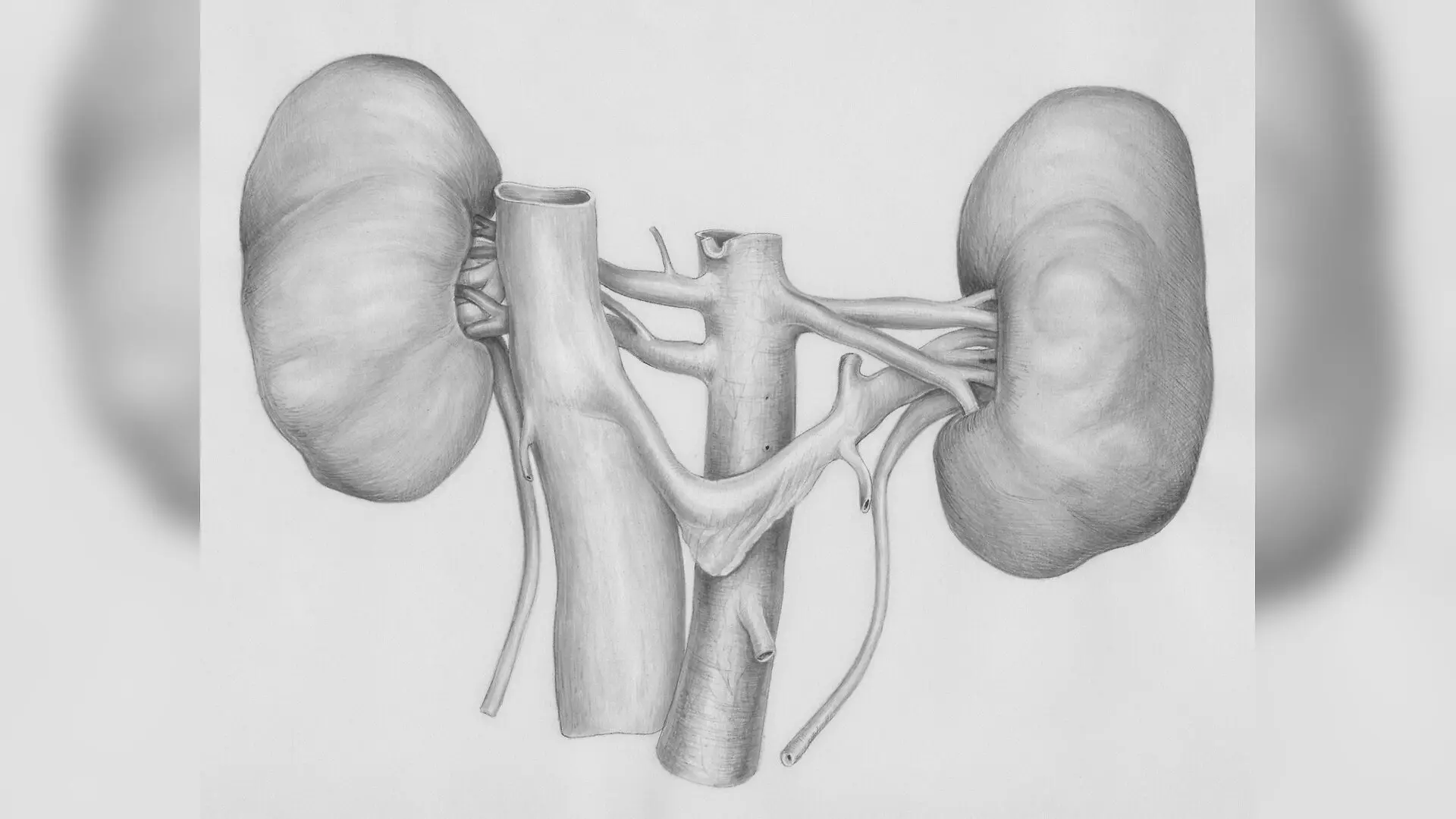
When oxygen levels plummet inside damaged kidneys, a molecular chain reaction begins that transforms healthy kidney cells into scar tissue and drives chronic kidney disease toward end stage failure. A team of researchers has uncovered how low oxygen conditions inside kidney tubules switch on a specific genetic pathway, shedding light on why some kidney injuries progress to permanent scarring.
How Low Oxygen Drives Permanent Kidney Scarring
Chronic kidney disease affects millions of people worldwide, and renal interstitial fibrosis, which is the excessive buildup of scar tissue in the spaces between kidney tubules, remains the ultimate pathway for advanced kidney failure. The study, published in Renal Failure, reveals that hypoxia, or oxygen starvation, acts as a primary trigger for this scarring process by activating a protein called hypoxia inducible factor 1 alpha (HIF 1 alpha).
To understand this mechanism, the authors examined human kidney biopsy samples from 5 patients diagnosed with renal interstitial fibrosis and 5 non fibrotic control subjects. They also tested an adenine induced chronic kidney disease rat model, where 6 week old male Sprague Dawley rats received daily doses of 250 milligrams per kilogram of adenine for three weeks to induce kidney failure.
The clinical and animal data confirmed that failing kidneys exhibited significantly elevated levels of blood urea nitrogen, serum creatinine, and prominent structural scarring shown through Masson trichrome staining. Immunohistochemical analysis revealed that both HIF 1 alpha and a transcription factor known as SOX9 were heavily upregulated in the renal tubular epithelial cells of fibrotic kidneys compared to healthy controls.
Laboratory Tests Reveal the HIF 1 Alpha and SOX9 Connection
To prove how hypoxia alters kidney cells, the researchers cultured rat renal tubular epithelial cells under hypoxic conditions containing 1 percent oxygen for up to 24 hours. While cell viability remained steady, the cells underwent a dramatic physical transformation, shifting from a normal cobblestone appearance into elongated, spindle shaped fibroblast like cells.
During this transformation, the expression of mesenchymal markers associated with scarring, specifically alpha smooth muscle actin and collagen type 1, increased significantly alongside rising levels of HIF 1 alpha and SOX9. When the team applied 2 methoxyestradiol, a pharmacological inhibitor of HIF 1 alpha, at concentrations ranging from 2 to 10 micromolars, it successfully blocked HIF 1 alpha production and subsequently suppressed SOX9 expression.
Conversely, when normoxic cells were treated with deferoxamine, an iron chelator that stabilizes HIF 1 alpha under normal oxygen conditions, it mimicked the hypoxic response. This chemical stabilization drove up SOX9 levels and triggered the expression of fibrosis markers without requiring actual oxygen deprivation.
What Happens When SOX9 Is Silenced in Kidney Cells
To determine if SOX9 is truly necessary for the scarring process, the researchers used small interfering RNA to silence SOX9 expression in cells exposed to hypoxia. When SOX9 was knocked down, the cells retained their normal epithelial shape and failed to develop the elongated, scar driving morphology typically caused by low oxygen.
Furthermore, silencing SOX9 reduced the accumulation of alpha smooth muscle actin and collagen type 1. These experiments demonstrate that HIF 1 alpha acts upstream to regulate SOX9, forming a functional signaling axis that directly promotes extracellular matrix deposition and tissue fibrosis.
Study Limitations and Future Therapeutic Horizons
While these findings map out a clear pathway for kidney fibrosis, the authors noted several limitations in their work. The clinical cohort included only a small sample of 5 patients with fibrosis and 5 controls, and the animal experiments relied exclusively on an adenine induced chronic kidney disease model rather than alternative models like ischemia reperfusion injury.
Additionally, the researchers did not investigate whether SOX9 might exert any feedback regulation on HIF 1 alpha, or map out every downstream target of SOX9 in the fibrotic cascade. Future research will need to test whether targeting the HIF 1 alpha and SOX9 axis can safely halt or reverse kidney scarring in living organisms.
The research was published in Renal failure on November 18, 2026.
This article has been fact checked for accuracy, with information verified against reputable sources. Learn more about us and our editorial process.
Last reviewed on .
Article history
- Latest version
Cite this page:
- Posted by David Anderson