How Migrating Cancer Cells Hijack Brain Neurons and Astrocytes to Form Deadly Secondary Tumors
A comprehensive review reveals how disseminated tumor cells actively reprogram the brain microenvironment to evade immune surveillance and drive metastatic outgrowth.

Brain metastasis develops when tumor cells from primary cancers like lung, breast, and melanoma successfully travel through the bloodstream and cross the blood-brain barrier to colonize neural tissue. A comprehensive review published in Cancer Communications by Laiba Anwar and colleagues at the University of Nebraska Medical Center reveals how these migrating cells actively remodel the brain microenvironment to survive, evade immune clearance, and eventually form deadly secondary tumors.
How Do Escaping Cancer Cells Cross the Blood-Brain Barrier?
The journey begins at the primary tumor, where growing cancer cells experience severe crowding, low oxygen, and high mechanical stress that drive local invasion and intravasation into circulation. While in the bloodstream, circulating tumor cells must endure fluid shear stress and immune surveillance before becoming mechanically trapped in narrow cerebral capillaries, particularly around capillary branch points and gray-white matter junctions. To breach the tightly sealed blood-brain barrier, these arrested cells release specialized extracellular vesicles and pro-invasive proteins such as matrix metalloproteinases that degrade endothelial junctions and facilitate active transmigration.
Once inside the brain parenchyma, metastatic cells rarely expand immediately into large growths. Instead, they often enter a prolonged state of dormancy within the perivascular niche, remaining quiescent for years or even decades before reactivation occurs. During this latent phase, successful survival depends heavily on reciprocal interactions with resident brain cells that dictate whether disseminated cells are eliminated or protected.
In What Ways Do Brain Cells and Immune Populations Aid Tumor Outgrowth?
Astrocytes, which are the most abundant glial cells in the central nervous system, act as early responders that can either eliminate invading cells or become co-opted to support tumor progression. Under sustained tumor signaling, reactive astrocytes undergo profound molecular changes, releasing cytokines and establishing gap junctions that transfer signaling molecules to cancer cells while dampening local immune responses. Microglia and infiltrating macrophages also accumulate around lesion margins, shifting toward an immunosuppressive state that fuels proliferation and shields the expanding tumor from immune attack.
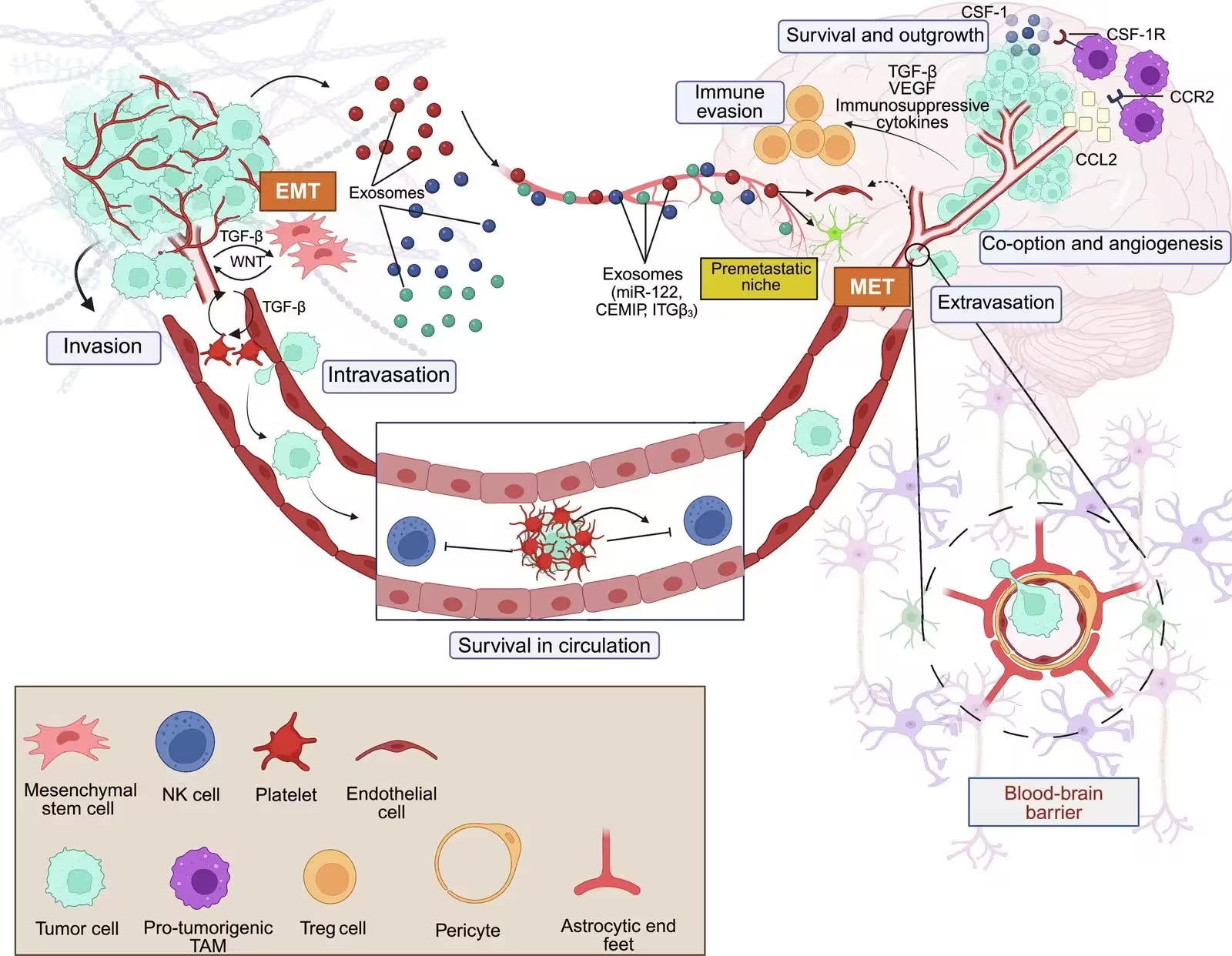
Beyond immune modulation, metastatic cancer cells engage in striking metabolic and neuronal adaptations to thrive in the nutrient-scarce brain environment. Research highlights how tumor cells hijack neuronal signaling pathways, forming functional synaptic connections and co-opting neurotransmitter networks to drive rapid growth. Some cancer lines absorb specialized substrates like lactate and glutamate through altered transporter expression, while others even acquire functional mitochondria directly from adjacent neurons to boost their energy metabolism and therapeutic resistance.
What Limitations Face Current Therapies and Future Approaches?
Despite aggressive treatment strategies including surgical resection, stereotactic radiosurgery, and whole-brain radiotherapy, overall survival for patients with brain metastases remains low. Systemic immunotherapies that prove effective against primary extracranial tumors frequently fail inside the brain due to the uniquely immunosuppressive microenvironment and restrictive vascular barriers. Addressing these challenges will require advanced artificial intelligence models for early diagnosis, spatial transcriptomics to map cellular heterogeneity, and combinatorial therapies that target both tumor survival signals and supporting stromal pathways.
The research was published in Cancer communications (London, England) on November 19, 2026.
This article has been fact checked for accuracy, with information verified against reputable sources. Learn more about us and our editorial process.
Last reviewed on .
Article history
- Latest version
Reference(s)
- Anwar, Laiba., et al. “Unraveling the Shaping of the Microenvironment in Brain Metastasis: From Dissemination to Overt Lesions.” Cancer Communications, vol. 46, November 19, 2026 American Association for the Advancement of Science (AAAS), doi: 10.34133/cancomm.0072. <https://doi.org/10.34133/cancomm.0072>.
Cite this page:
- Posted by Omar Mirza