
Normal Gastric Anatomy and Physiology
Thursday, 07 September 2017 20:21Anatomically, the stomach exhibits a division into four distinct parts: the cardia, fundus, body, and pyloric region. The cardia, situated at the uppermost portion, encircles the entrance of the esophagus and is characterized by its lining of mucus-secreting epithelium. In contrast, the epithelium of the fundus and the body of the stomach presents a diversity of cell types, including mucus-secreting cells, parietal cells responsible for the secretion of hydrochloric acid and intrinsic factor, and peptic cells, also known as chief cells, which release the proteolytic enzyme pepsinogen.
The pyloric part further divides into two segments: the pyloric antrum and the pyloric canal. This segment is distinguished by its lining, comprised of mucus-secreting cells alongside neuroendocrine cells known as G cells, which are responsible for the secretion of gastrin.

In the stomach, the process of breaking down ingested food occurs both mechanically and chemically, resulting in the formation of a semi-digested liquid known as chyme. Subsequently, upon relaxation of the pyloric sphincter, chyme proceeds into the duodenum.
Gastric acid secretion comprises three distinct phases: the cephalic, gastric, and intestinal phases.
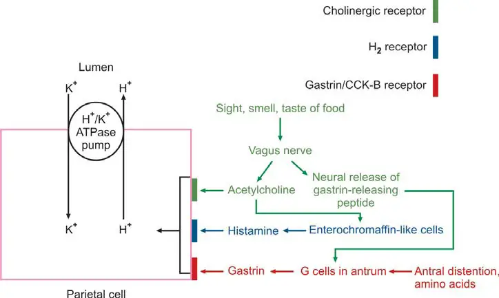
- Cephalic or Neurogenic Phase: This phase initiates upon sensory stimulation, triggered by the sight, smell, taste, or mere thought of food, leading to activation of vagal nuclei in the brain. Direct stimulation of parietal cells by the vagus nerve prompts acid secretion, while simultaneously stimulating antral G cells to release gastrin into the bloodstream, which further enhances gastric acid secretion. Notably, the cephalic phase is nullified by vagotomy.
- Gastric Phase: Initiated by the entry of ingested food into the stomach, the gastric phase ensues due to gastric distension. This distension, coupled with a rise in pH resulting from the neutralization of acid by food, triggers antral G cells to secrete gastrin into circulation. Gastrin, in turn, prompts the release of hydrochloric acid from parietal cells.
- Intestinal Phase: This phase occurs upon the entry of digested proteins into the duodenum, leading to an increase in stomach acid output. It is believed that certain hormones and absorbed amino acids play a role in stimulating parietal cells to secrete acid during this phase.
The collective secretion from the stomach is referred to as gastric juice, comprising several key constituents:
- Hydrochloric Acid (HCl): Secreted by parietal cells primarily located in the fundus and body of the stomach, HCl creates the highly acidic environment necessary for the activation of pepsinogen into pepsin. Gastric acid secretion is stimulated by histamine, acetylcholine, and gastrin. HCl serves to sterilize the stomach contents by eliminating most microorganisms and aids in the denaturation of proteins. Its secretion is inhibited by various factors including somatostatin, gastric inhibitory peptide, prostaglandin, and secretin.
- Pepsin: Produced by chief cells within the stomach, pepsin facilitates the partial digestion of proteins, resulting in the formation of large polypeptide molecules. Its optimal function occurs within the acidic pH range of 1.0 to 3.0, and its secretion is augmented by vagal stimulation.
- Mucus: Providing a protective barrier for the gastric mucosa, mucus helps prevent damage from stomach acid and digestive enzymes.
- Intrinsic Factor (IF): Essential for the absorption of vitamin B12 within the terminal ileum, IF is secreted by parietal cells of the stomach.

CONTRAINDICATIONS TO GASTRIC ANALYSIS
Thursday, 07 September 2017 18:53
LABORATORY TESTS FOR GASTRIC ANALYSIS
Thursday, 07 September 2017 18:53
Indications for Gastric Analysis
Thursday, 07 September 2017 18:18
Method of Gastric Analysis
Tuesday, 05 September 2017 18:51
Bioethics
Monday, 04 September 2017 13:27
Animal Biotechnology
Monday, 04 September 2017 13:17
Biophysics
Monday, 04 September 2017 13:09
Histopathology
Monday, 04 September 2017 12:55
Physiology
Monday, 04 September 2017 12:39
Food Science
Monday, 04 September 2017 12:26
Ecology
Monday, 04 September 2017 12:01
Forensic Science
Monday, 04 September 2017 11:36
Laryngospasm: Causes, symptoms, and treatments
Saturday, 02 September 2017 07:00
Microscopic Examination of Feces
Wednesday, 30 August 2017 18:26
CHEMICAL EXAMINATION OF FECES
Tuesday, 29 August 2017 20:21