Edition
EN
English
Login
Mode
BIOSCIENCE
Facebook
Twitter
Instagram
Menu
BSPK
Health Topics A-Z
Biology
Chemistry
Earth Science
Health
Medical Science
Physics
Space Science
More
Mode
Search
Menu
Search
Search
a
b
c
d
e
f
g
h
i
j
k
l
m
n
o
p
q
r
s
t
u
v
w
x
y
z
#
Cell Biology
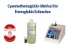
Cyanmethemoglobin (Hemoglobin Cyanide) Method for Estimation of Hemoglobin
How to Count Reticulocytes (Manual Method)
Mitochondria: Definition, Function, Structure and Facts
Red Cell Indices
Reticulocyte
Sahli’s Acid Hematin Method for the Estimation of Hemoglobin
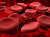
What is Hemoglobin? Types, Indications and Methods
Homepage
Login for unlimited free access
Login
Close the Menu
Explore by Subject
Astronomy
Biology
Biotechnology
Chemistry
Earth Science
Ecology
Environmental Science
Genetics
Health
Marine Science
Mathematics
Medicine
Physics
Psychology
Space Science
Technology
Urban Studies
Health Topics A-Z
Follow Us
Facebook
Twitter
RSS Feed
YouTube
Privacy Policy
Terms of Service
Contact Us
Copyright © 2012-2026 Dungrela Publishing