Coelom and Coelomoducts of Annelida
Coelom is a fluid-filled body cavity in Annelida that provides space for organ systems. Coelomoducts are excretory structures that remove waste products.
Coelom, a crucial feature of Annelida, is a fluid-filled body cavity providing space for organ systems and facilitates movement. Coelomoducts, found in some species, serve as excretory structures, eliminating metabolic waste products and maintaining osmotic balance. These structures contribute to the physiological functioning and overall complexity of the annelid body plan.
The space enclosed between the two mesodermal layers is called true coelom. The triploblastic animals are classified into three types based on the presence or absence of this coelom.
- Acoelomate: Coelom is absent. E.g.: Platyhelminthes.
- Pseudo coelomate: The blastocoel is retained in between the body wall and alimentary canal as pseudocoelom. E.g.: Nemathelminthes.
- Coelomate: True coelom is present. These are two types based on their formation.
- Schizo coelomate: By splitting of mesoderm layer this true coelom is formed. E.g.: Annelida, Arthropoda, and Mollusca.
- Enterocoelcmata: From the Archenteron of gastrula two mesodermal pouches will arise. They grow and unite to form true coelom. It is called enterocoelic coelome. E.g.: Echinodermata & chordata.
The coelom is filled with coelomic fluid. This coelomic fluid will perform the following functions in Annelid worms.
- It will keep all the body organs moist.
- It will allow the peristalasis of the alimentary canal.
- It works as a fluid skeleton and gives turgidity to the body.
- It will protect the body. Its phagocytic amebocyte will eat away the pathogenic bacteria which enter the body.
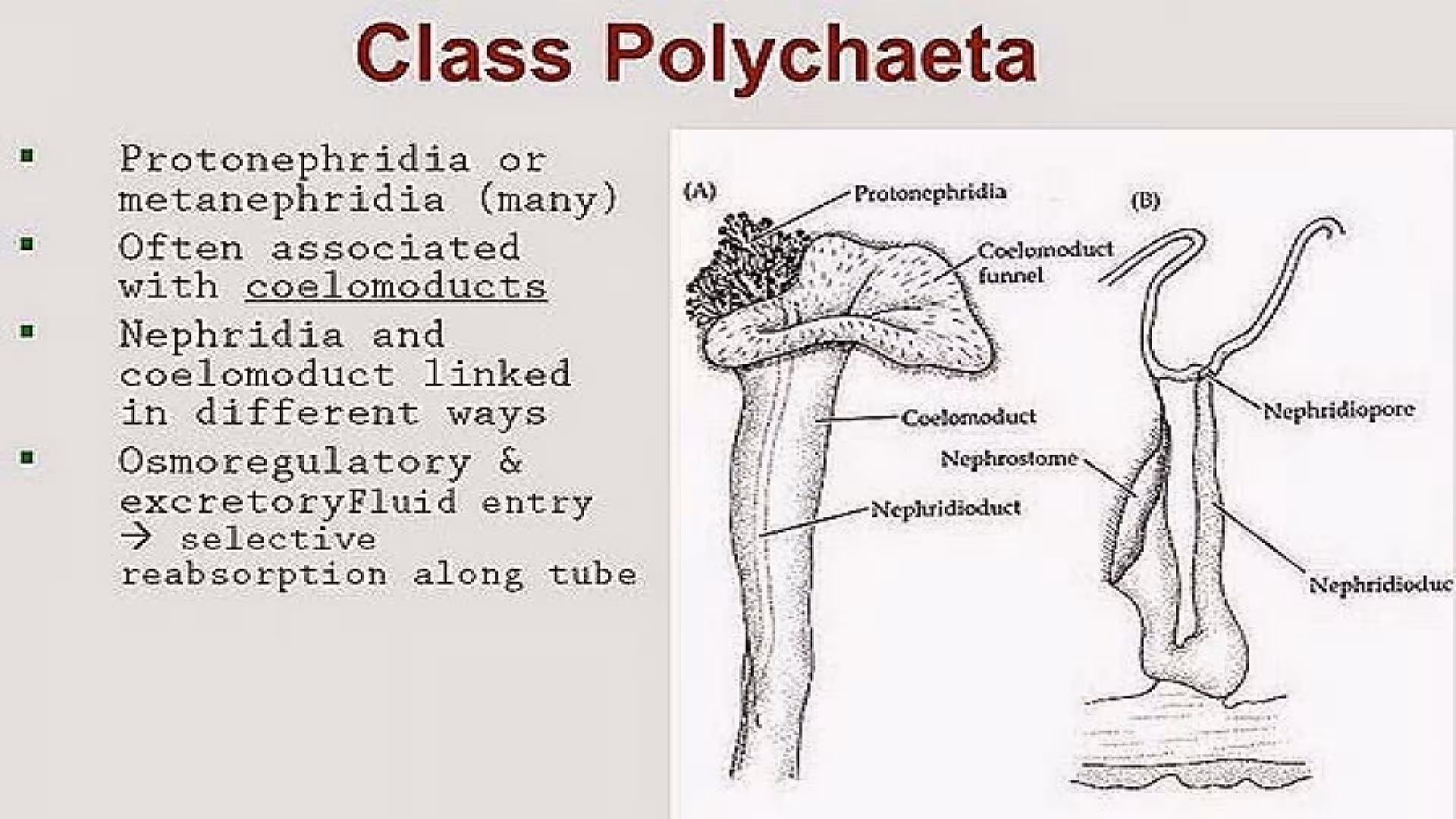
- Coelom ducts: In annelids, the body is divided into several segments. Each segment will show segmental structures. Coelom ducts and Nephridia are arranged segmentally in these organisms. They are derived from mesoderm. They are formed from coelom. They open out on the body through genital pores. They open into the coelom through ciliated funnels called coelomostomes. These coelom ducts work like genital ducts. Because of this, these coelomoducts are confined to only a few segments in some organisms they secondarily take up excretory function.
- Nephridia: These are derived from the ectoderm. They are excretory structures. They show cilia. They are arranged repeatedly in all segments. They open on the body wall through Nephridio pores. They open into the coelom through funnel-like nephrostomes. Different types of nephridia are present.
- Protonephridia: These are primitive nephridia. They end blindly in the coelom. They are seen in larval forms of polychaetes. They bear solenocytes. They resemble the flame cells of Platyhelminthes. These protonephridia are present in adults. Polychaetes like Vanadis, Tomopteris, etc.
- Metanephridia: They open into the body cavity through the nephrostome. It gives a small neck, it will pierce the septal wall and enter the next segment and coil these. There it opens on the body wall through the nephridiopore. Thus metanephridia will open on both ends. Such nephridia are seen in Neries, and members of polychaete, oligocheate, etc. If these nephridia open on the body wall they are called exo-nephridia. If they are connected with the alimentary canal, they are called enteronephric nephridia.
- Nephromixia: In Hirudinea, oligochaete and in some polychaetes nephridia and coelomoducts are separate. But in some polychaetes nephridia and coelomoducts unite to form nephromixia. Through these excretory matter and reproductive bodies are liberated. Different types of nephromixia are noticed.
- Protonephromixia: E.g.: Phyllodoce Protonephridia and coelomoducts are united.
- Meta nephromixia: E.g.: Hesione. Metanephridia and coelomoducts are united.
- Mixo-nephridium: E.g.: Arenicola. The coelomoduct and nephridium will unite as a single duct.
- Ciliated organs: E.g.: Neries. Coelom ducts are modified as ciliated organs. They are connected with longitudinal muscles and open on the body wall.
The information on this page is peer reviewed by a qualified editorial review board member. Learn more about us and our editorial process.
Last reviewed on .
Article history
- Latest version
Cite this page:
- Comment
- Posted by Dayyal Dungrela