Anatomy of Hirudinaria
Leech is an ectoparasite, which belongs to
- Phylum: Annelida
- Class: Hirudinea
- Order: Gnathobdellida
It has a dorsoventrally compressed body. It swims in the water in search of a suitable host.
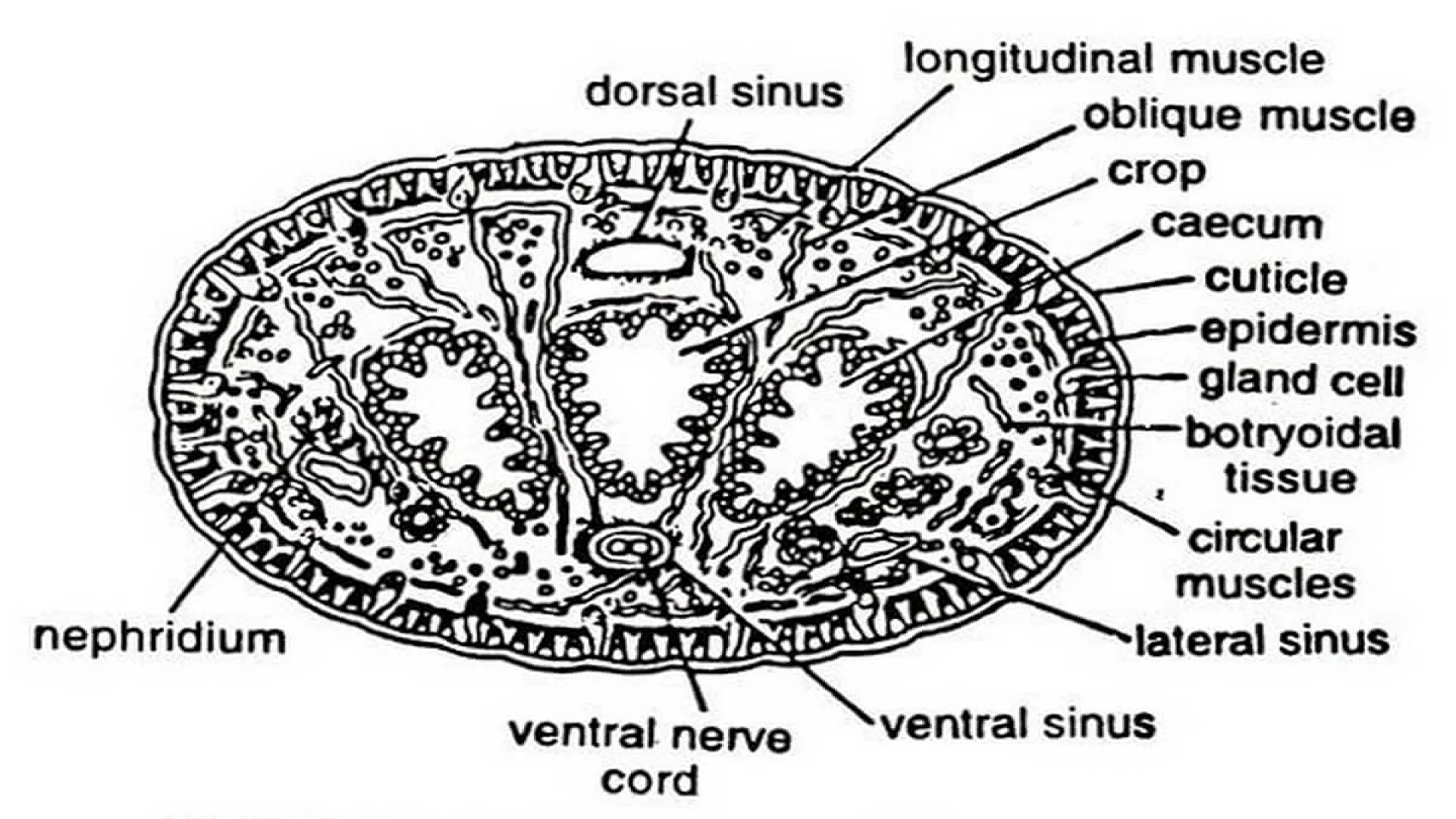
The transverse section of the leech shows the following parts.
- Dermo-muscular body wall.
- Botryoidal tissue.
- Organ system.
Dermo-muscular body wall
The body wall contains
- Cuticle
- Epidermis
- Dermis
- Muscular layer.
Cuticle
It is a thin, transparent, non-cellular covering of the body. It is secreted by the Epidermis. It is cast off from time to time.
Epidermis
It is single-layered. The cells are columnar. Their broader ends are placed to the outside. They are fitted together like a layer. The intercellular spaces present between the cells of the epidermis contain connective tissue, pigments, and haemocoelomic capillaries.
Epidermis shows glands and receptor organs.
Glands of the Epidermis
- Simple glands: They are present all over the epidermis. These glands are pear-shaped. They extend up to the dermis. They are nucleated. They open through a small opening on the epidermis. They produce mucous.
- Prostomial glands: In the Prostomium region the epidermis contains prostomial glands. They secrete the plugs of ootheca.
- Clitellar glands: The epidermis of the clitellum shows clitellar glands. They produce an egg case and albumen.
- Glands of sucker regions: Pre-oral and posterior suckers will show many epidermal glands. These glands produce mucous. Their secretion helps in locomotion and attachment to the substratum.
Receptors
In the epidermis many receptors are present.
- Eyes: In the first five segments, five pairs of eyes are present. (One pair in the first annulus of each segment)
- Annular receptors: In each annulus several receptors are present. They are sensitive to touch; (Tangoreceptors).
- Segmental receptors: In the first annulus of each segment bigger segmental receptors are present. They are tango receptors and photoreceptors.
- Dermis: It is present below the epidermis. It is made of fibrous connective tissue. It contains pigments, fat cells, muscle fibers, and haemocoelomic capillaries.
- Muscles: Below the dermis muscles are present.
- Circular muscles
- Oblique muscles
- Longitudinal muscles
- Dorso-ventral muscles
- Vertical muscles and
- Radial muscles
In the entire muscle group, the longitudinal muscle group is the biggest and most well-developed muscle. The muscles of the body will bring movements in the body.

Functions of the body wall
- It covers and protects the internal parts of the body.
- Receptors are helpful to locate their host and enemies.
- The muscles are helpful for locomotion.
- Secretions of the epidermal glands help in the formation of the cocoon.
Botryoidally tissue
In between the longitudinal muscles of the body wall and the alimentary canal, the cavity is filled with botryoidal tissue. The cell walls of this tissue have brown pigment. The tissue shows some spaces called haemocoelomic spaces. They are filled with haemocoelomic fluid.
Other organs:
In the cross-section, we can see the following parts enclosed in the botryoidal tissue.
- Dorsal haemocoelomic channel.
- Ventral haemocoelomic channel.
- Two lateral haemocoelomic channels.
- Alimentary canal.
- Ventral nerve cord surrounded by ventral haemocoelomic channel
- If the transverse section goes through 12 to 22 segments we can notice vas deferens, testis sac, and nephridium also.
The information on this page is peer reviewed by a qualified editorial review board member. Learn more about us and our editorial process.
Last reviewed on .
Article history
- Latest version
Cite this page:
- Comment
- Posted by Dayyal Dungrela