Stethoscope Anatomy: Understanding the Parts and Functions
Learn the essential parts of stethoscope anatomy and their functions, helping healthcare professionals make accurate diagnoses.
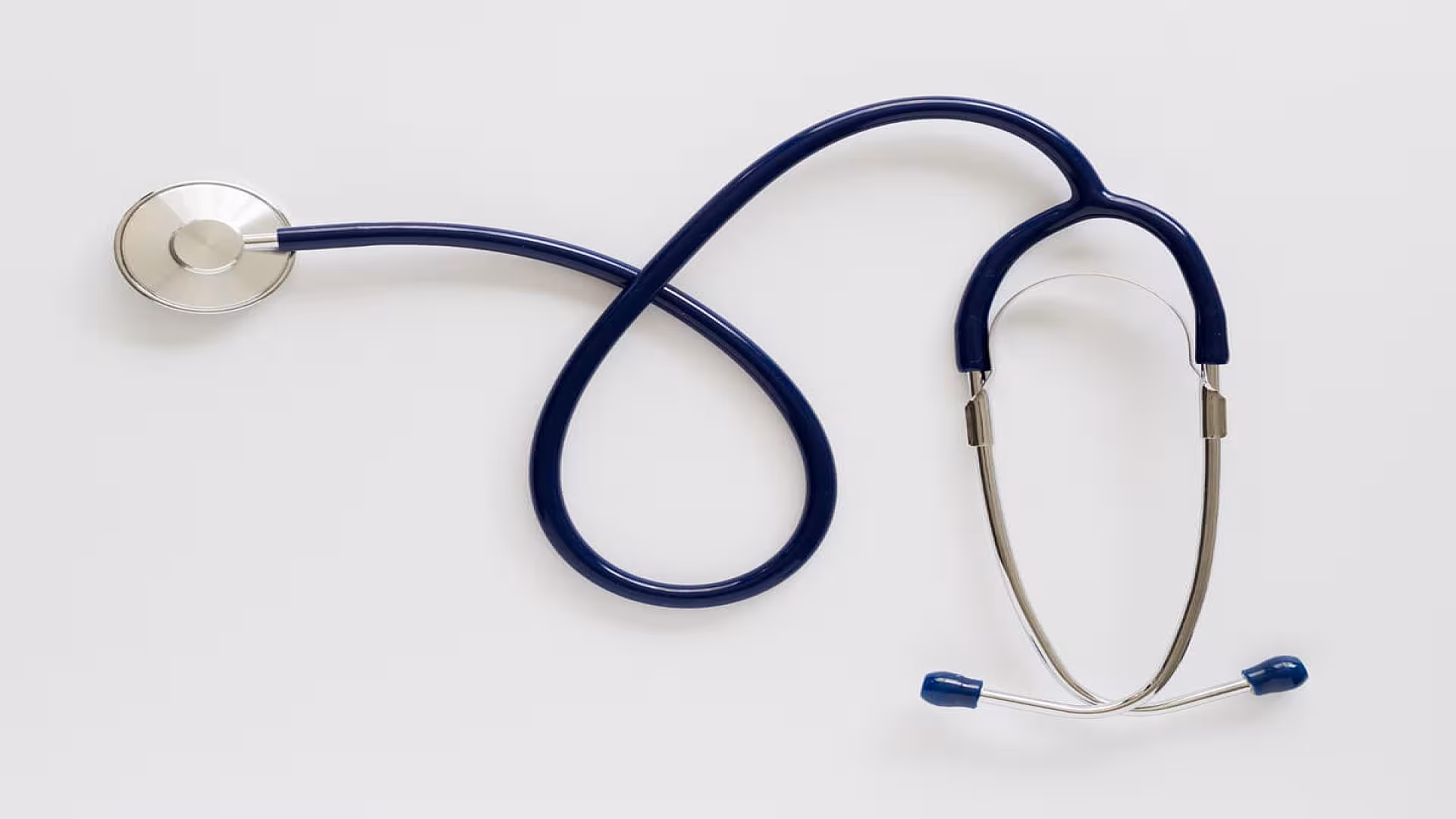
The stethoscope is an iconic tool in the medical field, instantly recognizable as a symbol of healthcare professionals. Despite its simple appearance, the stethoscope anatomy is complex and designed with precision to help detect and amplify sounds within the body.
The Chestpiece
At the heart of a stethoscope’s anatomy is the chestpiece, which comes into direct contact with the patient's body. The chestpiece houses two main components:
- Diaphragm: The larger, flat side of the chestpiece, used to hear high-frequency sounds like lung or heart sounds. It works by transmitting sound waves through the tubing to the listener's ears.
- Bell: The smaller, concave side, typically used to detect low-frequency sounds, such as certain heart murmurs or bowel sounds. Not all modern stethoscopes have a bell, as some feature a tunable diaphragm instead.
Tubing
The tubing in stethoscope anatomy plays a vital role in delivering sound from the chestpiece to the earpieces. Typically made from durable, flexible materials like PVC, the tubing is designed to maintain the integrity of the sound without interference from external noises. High-quality stethoscopes have thick, non-latex tubing to prevent sound leakage, ensuring clear and accurate acoustics.
The length of the tubing also matters. Standard stethoscopes have tubing lengths between 22 and 30 inches. Longer tubing may offer more comfort to the healthcare provider but can slightly diminish sound quality.
Headset
The headset connects the tubing to the earpieces and consists of three critical parts: the binaurals, the tension spring, and the earpieces.
- Binaurals: These are the metal tubes connected to the tubing. They are angled to fit comfortably in the user’s ears and direct sound efficiently. The binaurals are usually adjustable to accommodate different ear sizes.
- Tension Spring: Located inside the binaurals, the tension spring allows the user to adjust the tension for a snug fit. This feature enhances comfort during long hours of use.
- Earpieces: Soft, rubber tips that fit into the ears, sealing out ambient noise. They are designed to maximize comfort and sound clarity.
Diaphragm vs. Bell: Key Differences
Understanding the difference between the diaphragm and the bell is critical to using a stethoscope effectively. The diaphragm is most often used in routine check-ups as it is better suited for high-pitched sounds like normal heartbeats and lung sounds. In contrast, the bell is useful for hearing lower-pitched sounds, such as abnormal heart murmurs or bruits. For healthcare professionals, knowing when to use each part is essential for making accurate diagnoses.
Stethoscope Variations
While the basic stethoscope anatomy remains consistent, there are different types of stethoscopes designed for specialized purposes. For example:
- Cardiology Stethoscopes: These stethoscopes offer superior acoustic quality, allowing physicians to detect subtle heart sounds with greater clarity.
- Electronic Stethoscopes: Featuring amplification and noise cancellation technology, electronic stethoscopes are useful in noisy environments or for hearing faint sounds.
- Pediatric Stethoscopes: Designed with smaller chestpieces to accommodate infants and children, these stethoscopes provide better sound accuracy for pediatric patients.
Conclusion
A thorough understanding of stethoscope anatomy is fundamental for healthcare professionals who rely on this tool daily. From the diaphragm to the earpieces, each part of the stethoscope is carefully crafted to enhance sound clarity, making it possible to detect a wide range of bodily sounds. By mastering the use of a stethoscope, medical practitioners can make more accurate assessments, ultimately improving patient care.
In summary, stethoscope anatomy is more than just a technical subject; it’s about understanding how each part contributes to better diagnostic outcomes. Whether you're a seasoned professional or a medical student, mastering stethoscope usage will enhance your ability to provide effective healthcare.
Last reviewed on .
Article history
- Latest version
Cite this page:
- Posted by Dayyal Dungrela