Reticulocyte
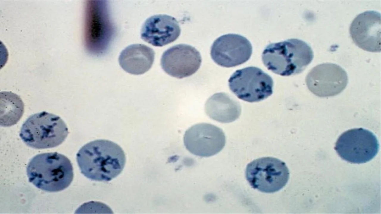
Reticulocytes are young or juvenile red cells released from the bone marrow into the bloodstream and that contain remnants of ribonucleic acid (RNA) and ribosomes but no nucleus.

Reticulocytes are young or juvenile red cells released from the bone marrow into the bloodstream and that contain remnants of ribonucleic acid (RNA) and ribosomes but no nucleus. After staining with a supravital dye such as new methylene blue, RNA appears as blue precipitating granules or filaments within the red cells. Following supravital staining, any nonnucleated red cell containing 2 or more granules of bluestained material is considered as a reticulocyte (The College of American Pathology). Supravital staining refers to staining of cells in a living state before they are killed by fixation or drying or with passage of time. Reticulocyte count is performed by manual method.
USES
- As one of the baseline studies in anemia with no obvious cause.
- To diagnose anemia due to ineffective erythropoiesis (premature destruction of red cell precursors in bone marrow seen in megaloblastic anemia and thalassemia) or due to decreased production of red cells: In hypoplastic anemia or in ineffective erythropoiesis, reticulocyte count is low as compared to the degree of anemia. Increased erythropoiesis (e.g. in hemolytic anemia, blood loss, or specific treatment of nutritional anemia) is associated with increased reticulocyte count. Thus reticulocyte count is used to differentiate hypoproliferative anemia from hyperproliferative anemia.
- To assess response to specific therapy in iron deficiency and megaloblastic anemias.
- To assess response to erythropoietin therapy in anemia of chronic renal failure.
- To follow the course of bone marrow transplantation for engraftment.
- To assess recovery from myelosuppressive therapy.
- To assess anemia in neonate.
The information on this page is peer reviewed by a qualified editorial review board member. Learn more about us and our editorial process.
Last reviewed on .
Article history
- Latest version
Cite this page:
- Posted by Dayyal Dungrela