AI Reads Laser Light Pulses to Spot Hidden Malaria Parasites
A new deep learning approach bypasses the need for fluorescent tags by analyzing how individual cells scatter laser light.
Invisible malaria parasites hiding inside mosquito tissue can now be spotted in real time by an artificial intelligence trained to read laser light pulses. Researchers have developed a label-free detection method that separates rare infectious organisms from background cellular debris without needing fluorescent chemical tags or genetic modifications.
Flow cytometry is a standard laboratory technique used to analyze thousands of individual cells per second by passing them through a focused laser beam. While traditional cytometers rely heavily on fluorescent dyes or engineered glowing reporter proteins to identify specific targets, finding rare biological particles in complex mixtures remains a significant bottleneck. In malaria research, isolating the infectious stage of the parasite from mosquito salivary glands typically yields a chaotic mixture dominated by insect tissue and cellular fragments.
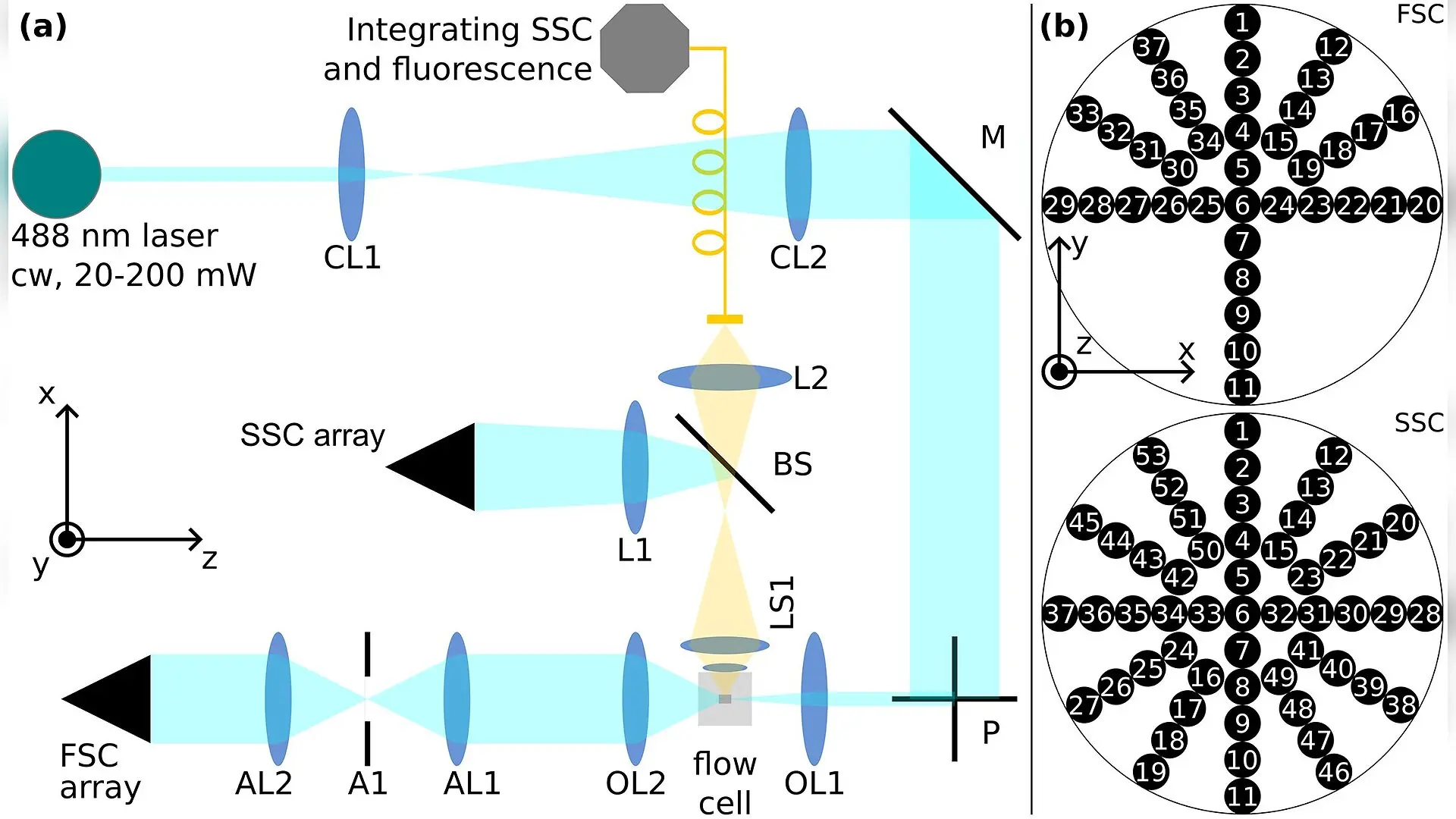
To overcome this hurdle, researchers turned to a specialized technique called multi-angle pulse shape flow cytometry, which captures the temporal dynamics of scattered laser light across multiple distinct angles rather than just measuring total light intensity. Because biological particles have unique shapes and internal structures, the way they scatter light as they travel through a laser beam creates a distinct electrical fingerprint. The study was published in PLOS One on November 5, 2026, detailing how machine learning can interpret these subtle optical signatures.
How Laser Scattering Reveals Hidden Parasites
When an object flows through the optical setup, the instrument records complete signal pulses across seven different light scatter channels at a sampling rate of 25 megahertz. Plasmodium falciparum sporozoites, which measure just a few micrometers in length, align themselves along the direction of fluid flow due to hydrodynamic forces. This consistent orientation produces reproducible pulse shapes that differ markedly from irregular mosquito cell fragments.
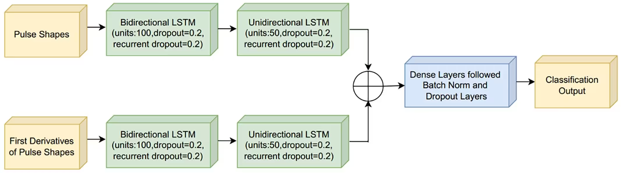
To process these complex temporal waveforms, the research team implemented a deep neural network architecture combining bidirectional long short-term memory layers with standard unidirectional recurrent layers. This design allows the algorithm to analyze both the raw pulse profiles and their first derivatives simultaneously, capturing how light intensity changes over time as a particle traverses the laser focus. By tracking these microsecond-scale fluctuations, the network learns to differentiate between true parasite signals and background noise.
Overcoming Class Imbalance with Artificial Intelligence
Working with biological samples where target organisms make up less than one percent of total events presents a severe statistical challenge for machine learning models. Early tests using models trained on single experimental datasets revealed high variability, as day-to-day instrument fluctuations and biological diversity caused performance to drop when applied to unseen test sets. To build robustness, the authors merged training data collected across three separate days, exposing the neural network to a wider range of experimental variation.
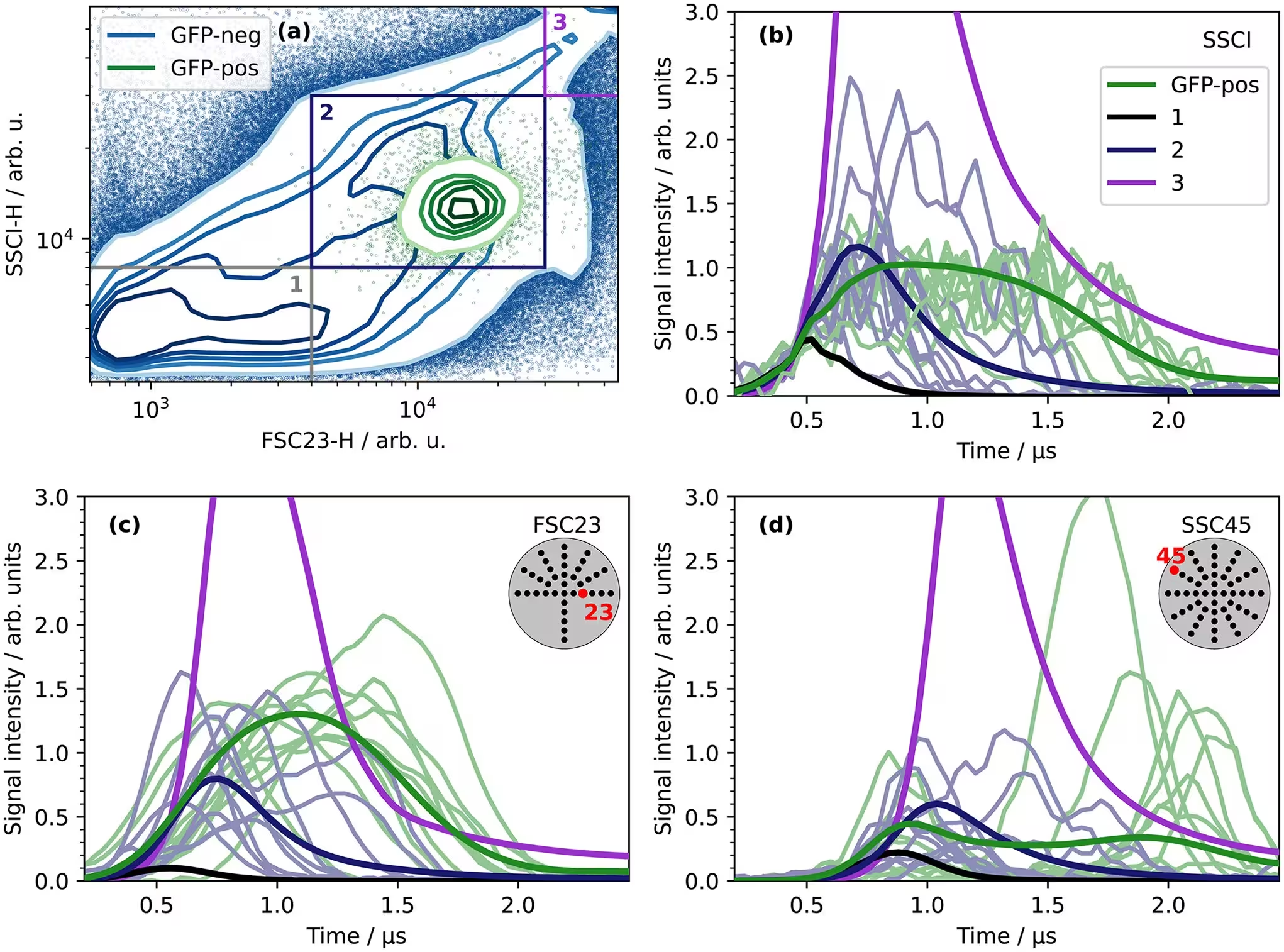
The team also applied a confidence threshold of 98 percent to the model predictions, filtering out ambiguous events where the classification probability fell below the cutoff. When tested on independent datasets containing roughly one million events each, this thresholding strategy dramatically improved classification precision from 11.35 percent up to 60.23 percent while retaining a parasite detection rate of nearly 99.9 percent.
Potential Limitations and Future Applications
Despite the high accuracy achieved with merged training sets, microscopic analysis of misclassified events pointed to intriguing biological complexities. Some non-parasite events that triggered false positive alarms exhibited pulse shapes nearly identical to true sporozoites. The authors suggest these particles may represent damaged or degraded parasites that lost their fluorescent reporter proteins but retained their elongated morphology, meaning some apparent algorithm errors might actually stem from limitations in manual ground-truth labeling.
Current sorting hardware cannot yet run complex deep learning inferences in real time to physically isolate live parasites during a sort. However, the authors note that integrating these neural network architectures into high-speed sorting platforms could soon enable label-free isolation of wild-type malaria parasites at scale, bypassing the need for genetically modified fluorescent strains and accelerating future vaccine and drug research.
The research was published in PLOS One on November 5, 2026.
This article has been fact checked for accuracy, with information verified against reputable sources. Learn more about us and our editorial process.
Last reviewed on .
Article history
- Latest version
Reference(s)
- Devaraj, Vinod., et al. “Identification of Plasmodium sporozoites using angle-resolved scattered light pulse shapes.” PLOS One, vol. 21, no. 11, November 5, 2026, pp. e0354060 Public Library of Science (PLoS), doi: 10.1371/journal.pone.0354060. <https://doi.org/10.1371/journal.pone.0354060>.
Cite this page:
- Posted by Farah Siddiqui