New Screening Test Reveals How Brain Thinning Maps To Memory Loss
A novel cognitive assessment for older adults successfully tracks physical changes in brain regions vulnerable to Alzheimer’s disease.
A new cognitive screening test developed in Latvia reveals a striking parallel between a person’s mental test scores and the physical thinning of their brain, offering fresh hope for spotting early signs of neurological decline. Researchers tested 106 older adults with varying degrees of memory and thinking abilities and discovered that scores on the newly devised assessment reliably track the structural health of key brain regions linked to Alzheimer’s disease.
Testing Memory Against Brain Structure
Medical experts face a growing challenge as populations age and cognitive disorders become more prevalent. Traditional screening tools often struggle with cultural biases or fail to capture subtle early shifts in reasoning and memory. To address these gaps, researchers designed the Riga Cognitive Screening Task, known as RiTa, which blends lifestyle history with specific mental challenges.
The research team set out to measure the concurrent criterion validity of the screening tool, checking whether individuals who struggle on the test also show physical changes in brain regions vulnerable to aging. The study, published in PLOS One on November 19, 2026, involved participants ranging in age from 55 to 92 years old. Among the 106 participants, 39 had normal cognition, 47 had mild cognitive impairment, and 20 had been diagnosed with Alzheimer’s disease.
Every participant underwent a 15 to 20 minute cognitive assessment alongside a detailed brain scan using a 3.0 Tesla magnetic resonance imaging scanner. The scans allowed scientists to measure the cortical thickness of 18 specific regions in the temporal and parietal lobes of the brain, areas known to suffer early damage during neurodegenerative processes.
How Multivariate Analysis Links Mind and Brain
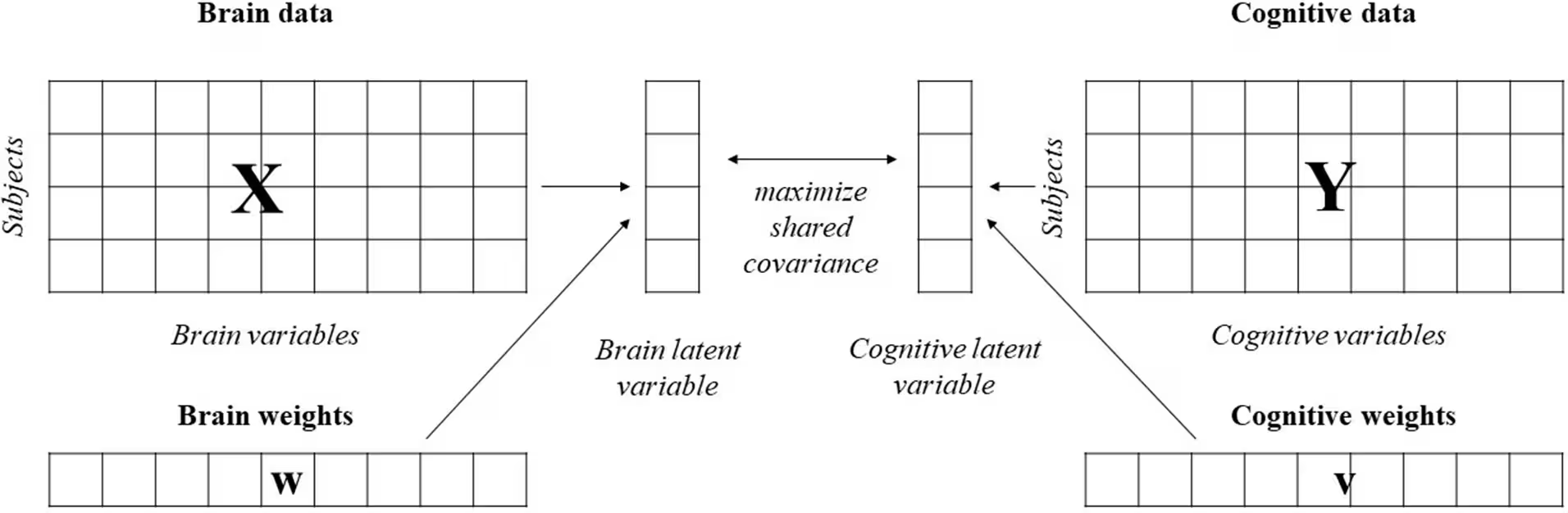
To analyze the complex relationship between brain anatomy and cognitive test scores, the authors used a statistical method called behavioural partial least squares correlation. This technique examines how two large sets of data, in this case brain measurements and cognitive performance scores, covary together as a unified system rather than looking at isolated pairs of variables.
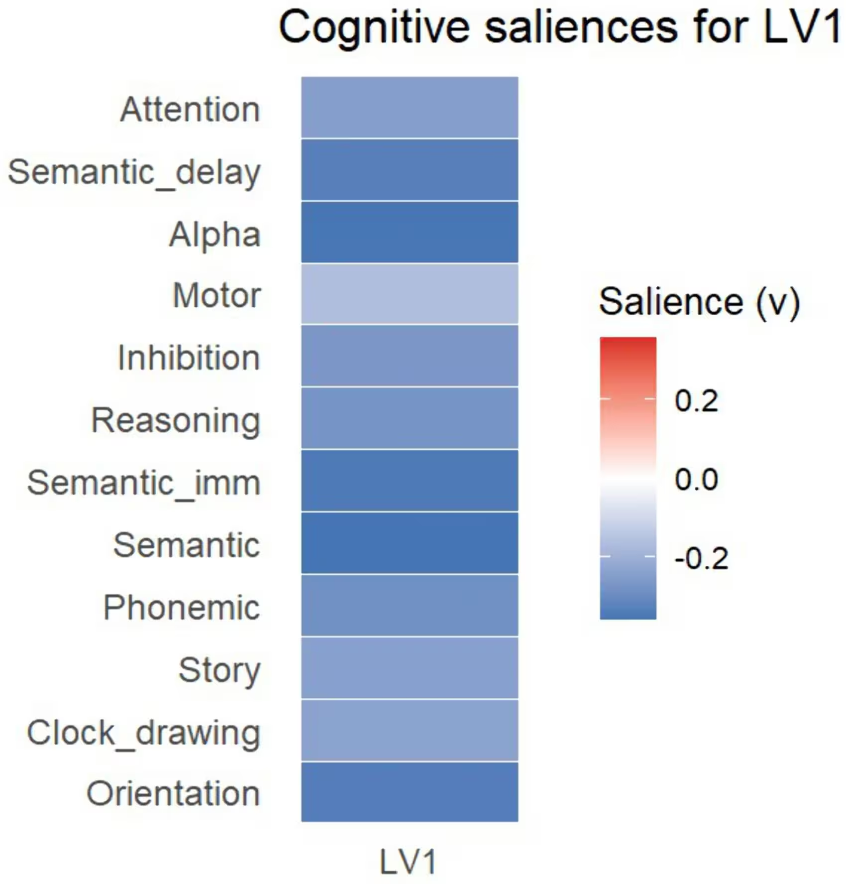
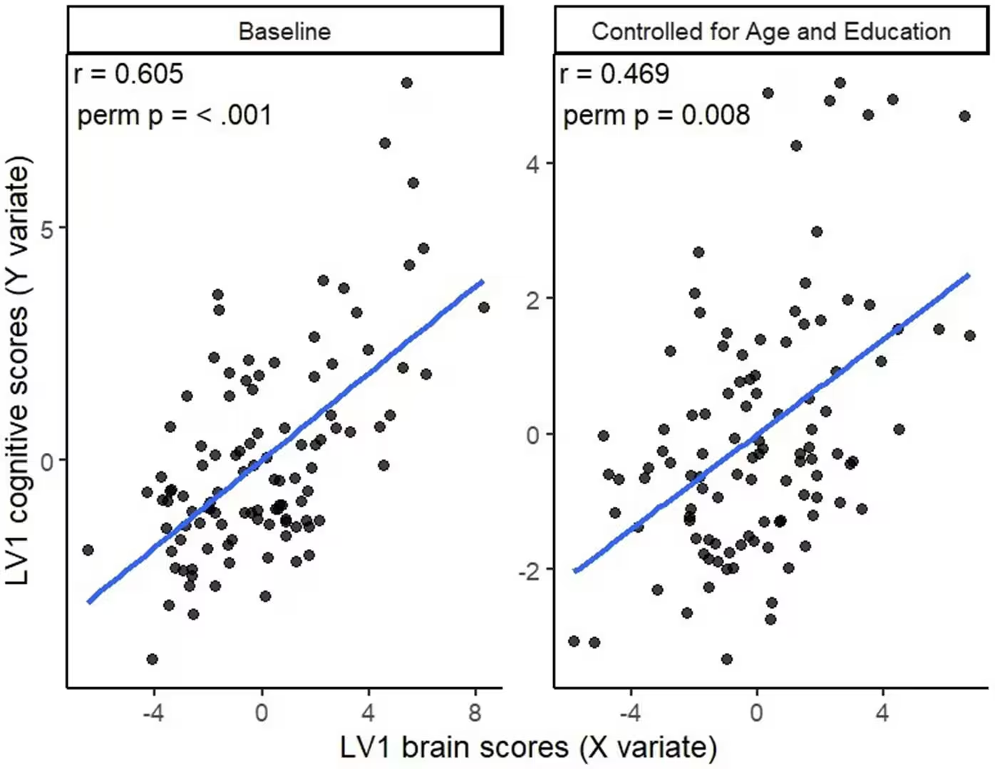
The analysis revealed a single statistically significant latent variable that captured a strong association between brain thickness and cognitive performance, yielding a correlation of 0.605. The primary contributors on the brain side included the entorhinal cortex, parahippocampal gyrus, middle temporal cortex, inferior parietal lobule, and supramarginal gyrus in both hemispheres. On the cognitive side, core tasks involving memory recall, working memory, orientation, verbal fluency, and executive inhibition drove the pattern.
When the researchers controlled for the confounding effects of age and education, the correlation between brain thickness and test scores dipped to 0.469 but remained statistically significant. This adjustment demonstrated that while age and schooling contribute to the observed relationship, they do not fully explain the deep structural connection between test results and cortical health.
What These Findings Mean for Clinical Practice
The results indicate that lower performance across memory, language, and executive tasks on the RiTa screening tool covaries with greater physical thinning in temporal and parietal cortical areas. These specific brain regions play vital roles in memory representation, contextual integration, and associative processing. By bypassing rigid divisions between clinical patients and healthy controls, the study captured a broad continuum of cognitive function.
The authors acknowledge several limitations in their work, including its cross-sectional design, which captures only a single snapshot in time. Because the study was not longitudinal, it cannot prove the exact chronological sequence of whether cortical thinning precedes cognitive decline or vice versa. Additionally, the recruitment method relied on senior organizations and clinics, meaning the sample may not represent the general population of older adults in Latvia.
Future research will need to follow participants over time to track whether changes in RiTa scores can predict progressive brain atrophy. External replication in independent samples will also help confirm the stability of these neurocognitive patterns, ultimately moving clinical practice closer to more accurate, early detection of cognitive impairment.
The research was published in PLOS One on November 19, 2026.
This article has been fact checked for accuracy, with information verified against reputable sources. Learn more about us and our editorial process.
Last reviewed on .
Article history
- Latest version
Reference(s)
- Šneidere-Pītersa, Kristīne., et al. “Multivariate cortical thickness-cognition covariance supports the criterion validity of the Riga Cognitive Screening Task.” PLOS One, vol. 21, no. 11, November 19, 2026, pp. e0344718 Public Library of Science (PLoS), doi: 10.1371/journal.pone.0344718. <https://doi.org/10.1371/journal.pone.0344718>.
Cite this page:
- Posted by Rabia Shah