Displaying items by tag: Article
Tuesday, 04 July 2017 12:31
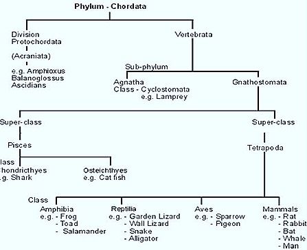
CLASSIFICATION PHYLUM CHORDATE

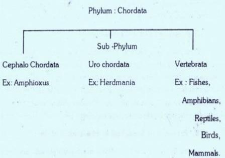
Phylum Chordate is divided into three Sub-Phyla.
- SUB-PHYLUM: UROCHORDATA
- SUB-PHYLUM: CEPHALOCHORDATA
- SUB-PHYLUM: VERTEBRATA
The first two sub phyla are called lower chordates or PROTOCHORDATES. They are usually called Acrania group. The vertebrata sub-phylum is called CRANIATA group.
1. SUB-PHYLUM: UROCHORDATA:
The urochordate animals belonging to this sub-phylum re called sea-squirts. The adults are marine and mostly fixed. Free living forms are also present. The body is covered by test or tunicin. The notochord and nerve cord are found in the larval stage. in the adult form they are completely lost. Gill-slits may be permanent. They are’ bisexual forms. The larvae are free swimming.
Ex: Olkopleura, A.scidian, Herdmania, etc.
2. SUB-PHYLUM: CEPHALOCHORDATA:
Cephalochordata are marine organisms. Their body is fish-like with notochord and nerve cord persisting throughout life . They extend the entire length of the body. The eyes, and jaws are absent. The fundamental plan of the chordate body is seen in its most simple form in these animals. Gonads are paired in Amphioxus and unpaired in Asymmetron.
Ex: Amphioxus, Asymmetron.
3. SUB-PHYLUM: VERTEBRATA OR CRANIATA:
Vertebrata are well developed chordates. They show distinct head. Notochord is replaced by the vertebral column completely or partially. The nerve-tubes anterior and enlarged to form a brain. Cranium protects the brain. Visceral clefts called Gills perform respiration. They are not more than seven pairs. Heart is ventral Andes are present They are formed by several segments.
Published in
Zoology
Tagged under
Tuesday, 04 July 2017 11:43
BIOLOGY OF CHORDATES

CHORDATES
The Phylum Chordate includes bilaterally symmetrical, metamerically segmented, triploblastic, enterocoelomate metazoans exhibiting hights complex organization. This phylum is supposed to be one of the most heterogenous and diversified gruop of animal kingdom. The minute sessile cephalodiscus, work-like Balanoglossus, degenerate tunicates, Amphioxus and the true vertebrates have been included in this phylum.
The vertebrates are fishes, amphibians, reptiles, birds and mammals. All these animals appear to be quite different from each other .However all of them possess certain common characters. These common characters are the presence of Notochord, dorsal nerve cord and gill-slits.
PRIMARY CHARACTERS OF CHORDATE
1. Presence of notochord or chorda dorsalis:
All Chordates porsess a solid, un segmented and flexible axial rod extending the whole length of the body. It is present mid dorsally and immediately above the alimentary canal and below the dorsal nerve cord. It porsess large vacuolated parenchymatous cells and enclosed in an inner elastic sheath and outer chordal sheath of dense fibrous connective tissue.
The notochord serves s a primitive internal skeleton. It also supports the central nervous system and the segmented muscles. In vertebrates the notochord is completely replaced by the vertebral column.
2. Presence of dorsal nerve cord:
The nervous system of chordates is the form of hollow tube present middorsally above the notochord and below the body wall. It is ectodermal in origin. From the ectoderm a medullary groove is formed which lateral develops neural tolds from its edges and forms the neural tube. Its cavity is known as Neurocoel. In higher chordates the anterior part of the neural tube develops into brain and the remaining part is called spinal cord.
3. Presence of Branchial or Pharyngeal clefts:
In the life cycle of all chordates the branchial clefts are found universally at some stage. These are formed as perforations in the laterally of pharynx and open to the exterior. Such gill-clefts are developed in every chordate. In certain animals (Amphioxus) these remain throughout life. In Aquatic chordates (fishes) the visceral clefts develop vascular Lamellae gills. These clefts are known as gill-clefts or gill-slits.
In terrestrial chordates these are seen during early development but in the adults modified into lungs. In the lows chordates (hemichordate, Cephalochordate & fishes) visceral clefts help in feeding as well as assist in respiration. In certain terrestrial forms these re modified into endocrine glands.
Origin of Chordates
There is no reliable evidence in favor of chordate origin. Origin of chordates has no definite conclusions because of lack of fossil evidences.
Recent investigations reveal the relationship among echinoderms, hemichordates and chordates. Certain phosphate is reported in all the three groups which is useful for muscle contraction Echinoderms and Hemichordates are developed from the common ancestor. So the chordates might have developed from the hemichordates.
The phylum chordata is classified into three sub-phyla. (Hemichordate has been given separate phylum status recently).
Simpler forms of chordate animals with notochord completely or incompletely formed are known as protochordates. These sub-phyla cephalochordata, urochordata (formerly Hemichordate) animals may resemble the ancestors of chordates. The vertebral column and skull are not formed in these animals.
In higher chordata animals like vertebrates animals central nervous system with brain & spinal cord, vertebral column and skull are formed along with other respective systems.
Published in
Zoology
Tagged under
Tuesday, 04 July 2017 11:28
GENERAL CHARACTERS OF CHORDATES

The animal kingdom is divided into two sub-kingdoms:
- Chordata
- Non-Chordata
GENERAL CHARACTRS OF CHORDATES
The Chordate characters appear in the developmental stage will persist in the adult or modified in the adult or disappeared in the adult
They are,
1. PRESENCE OF NOTOCHORD: It is an elastic, longitudinal stiff rod present between the nerve cord and alimentary canal. It is covered by an outer chodal sheath and inner elastic sheath or elastic-internal. Below it vacuolated, non-nucleated cells are present.
Notochord is present in the embryos of the vertebrates, but is replaced by vertebral column. In less-developed vertebrates the notochord is present throughout the life.
2. PRESENCE OF PHARYNGEAL GILL SLITS: Gill slits are present on either side of the pharynx. Each gill slit develops in the embryonic stage by evaginations of endoderm in pharynx with a corresponding invagination of ectoderm on the outside of the body. They are useful for respiration. In reptiles, birds and mammals, there are several pairs of gil slits in embryonic life, but they are not functional hence they are closed.
3. PRESENCE OF DORSAL TUBULAR NERVE CORD: The nerve cord in chordates is a hollow tube, situated dorsal to the alimentary canal and the notochord. It develops from the ectoderm. The cavity of nerve cord is called neurocoel.
These chordate characters appear in the developmental stages and they may remain or change or disappear in the adult.
The following characters are seen in all most all chordates.
4. TRLPLOBLASTIC NATURE: They possess three germ layers, 1. Ectoderm, 2. Mesoderm, 3. Endoderm.
5. DEVELOPMENT OF TRUE COELOME: In all the chordates a true coelome is present. It develops from mesoderm. The coelome is developed by enterocoelic method.
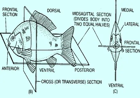
6. PRESENCE OF BILATERAL SYMMETRY: The body of the chordates show bilateral symmetry. Their body can be divided into two equal halves, through a sagittal plane which passé longitudinally.
No chordate animal possesses ideal bilateral symmetry, but they are near to such condition.
7. REDUCTION OF METAMERIC SEGMENTATION: Among the chordates, metamerism is seen in the internal structures. The myomeres are seen in lower chordates and segmentation is seen in the embryonic condition of higher vertebrates.
Published in
Zoology
Tagged under
Tuesday, 04 July 2017 11:12
ORIGIN OF CHORDATES

THEORIES ABOUT ORIGIN OF CHORDATES
It is believed that Chordates have originated from invertebrates. It is difficult to determine from which invertebrate group the chordates were developed. Chordate ancestors were soft bodied animals. Hence they were not preserved as Fossils.
Many biology theories were put forward to explain the Origin of Chordates.
1. COELENTERATE THEORY - ORIGIN OF CHORDATES: According to this theory chordates were developed from coelenterates. Radial symmetry coelenteron,cnidoblasts etc, were 1st and advanced characters were developed to give rise to chordates. This theory infers that chordates might have acquired higher characters independently. It is not correct and hence this theory is not acceptable.
2. ANNELID THEORY - ORIGIN OF CHORDATES: This theory suggests that the chordates have evolved from an annelid stock. The annelids show bilateral symmetry, mesmerism, head, lateral Coelome complete digestive tract, closed circulatory system, hemoglobin, etc., like chordates. The resemblance is enhanced if, an annelid is turned upside down. But the mouth would be dorsal which is unlike that of chordates. Metamerism and appendages of annelids differ in nature from those of the chordates. Bilateral symmetry, head and complete digestive tract occur in other non-chordate phyla also. Coelome is schizocoelic in annelids and enterocoelic in lower chordates. Haemoglobin is dissolved in the plasma in annelids but it is present in the red blood corpuscles in chordates. Annelid nerve cord is double, and, ventral in contrast to single, hollow, dorsal nerve cord of chordates. Some striking differences exist between the annelids and the chordates in their embryology. Hence it is difficult to accept this theory.
3. ECHINODERM - HEMICHORDATE THEORY - ORIGIN OF CHORDATES: This theory infers origin of chordates, hemichordates and echinoderms from a common ancestor. This theory is based on the following evidences.
a) EMBRYOLOGICAL EVIDENCE - ORIGIN OF CHORDATES: Both echinoderms and chordates have enterocoelic coelome, mesoderm and deuterostomous mouth. There is resemblance between the bipinnaria larva of certain echinoderms and the tornaria larva of hemichordates. In echinoderms chordates the central nervous system develops from a dorsal strip of ectoderm.
b) SEROGICAL EVIDENCE - ORIGIN OF CHORDATES: A close similarity exists between the proteins of the body-fluid of chordates and echinoderms. Hence the chordates are more related to echinoderms.
The radial symmetry of adult echinoderms will disprove their relationship with the bilaterally symmetrical chordates. In echinoderms radial symmetry is secondarily developed from a basically bilateral symmetry. Both the primitive and the early echinoderm larvae show bilateral symmetry.
The generalised chordate ancestry is as follows: This was suggested by N.J.Berrill, (1953) in his book “The origin of Vertebrates”.
ECHINODERM - AURICULARIA - HEMICHORDATA - TORNARIA - PROTO CHORDATE - ASCIDIAN TADPOLE - FREE SWIMMING CHORDATE
Hyman (1959) and others concluded that all the three groups have a common ancestor, Echinoderms and hemichordates branched off from a common evolutionary line which ended in the chordate group.
Published in
Zoology
Tagged under
Monday, 03 July 2017 10:54
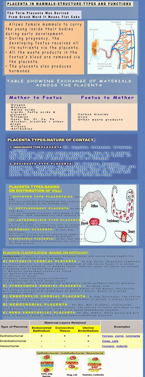
PLACENTA IN MAMMALS: STRUCTURE, TYPES AND FUNCTIONS

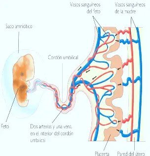
In Eutherian mammals the embryo develops in the uterus of mother. The developing embryo will get nourishment from mother through the placenta. Placenta is not common to all mammals. It is developed well in Eutheria The term placenta was delved from Greek word it means flat cake. Placenta is a special connective tissue, which contains the uterus of mother and foetal membranes of foetus.
Prototherian mammals are egg laying mammals. Hence placenta is not formed in uterus.
In marsupials the embryo develops incompletely in the uterus. They show yolk sac placenta and primitive allantoic placenta: Yolk sac placenta:
Ex: Diadelphis.
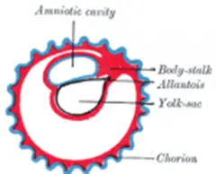
In these animals the developing embryo shows small allantois. It will never come in contact with chorion. Their yolk sac was large. It comes in contact with chorin. This part will gain blood vessels. This part will come in contact with endometrium of uterus. This is only a contact, but not fusion. Through this contact the embryo will absorb nourishment from mother. This is called chorio vitelline placenta or Yolk sac placenta.
Primitve Allantoic Placenta:
In paramoles simple allantoic placenta is developed Allantoic will enlarge It comes In contact with chonon This structure will be closely applied to mother’s uterus. It is called chorio-allantoic placenta. In these animals yolk sac placenta is not seen.
Placenta In Eutherla:
In Eutherian mammals true allantoic placenta is seen. Allantoic becomes big and comes in contact with chorion. This part will show close association with uterine wall. This connection is called placental connection. The structure of placenta will vary in different orders of Eutheria.
Placenta is classified in three ways.
- The placenta classification on nature of contact.
- Placenta is classified basis on the distribution of villi.
- Classification of placenta basing on histology.
NATURE OF CONTACT:
It is two types, Indeciduate and deciduate type.
Indeciduate type placenta: Ex: Ungulate, Cetaceans, Sirenians. Lemurs:
The chorianic villi are simple projections, they lie in contact with uterus. They have a loose contact. There is no fusion. At the time of birth of embryo uterus is not damaged.
Deciduate type Placenta: Ex: Primates, Rodentia, Insectivora, chiroptera The allantochorianic villi penetrate into uterine valli. They are intimately fused. Hence at the time of birth, the uterus is damaged. Bleeding occurs, the utrine wall enters into formation of placenta is called deciduas.
BASING ON DISTRIBUTION OF VILLI:
According to the distribution of villi five kinds of placenta are seen.
- Diffused type placenta: Ex: Horse, pig. The villi are uniformly distributed on the surface of blastocyst, except at the extreme ends.
- Cotyledonary placenta: Ex: Sheep, Cow, Deer. The villi are arranged in groups. Each group is called cotyledon. Each cotyledon fits into caruncla fo uterus.
- Intermediate type Placenta: Ex: Cainel, Giraffe. It is a rare type, it shows free villi on cotyledons. Hence it is called intermediate type placenta.
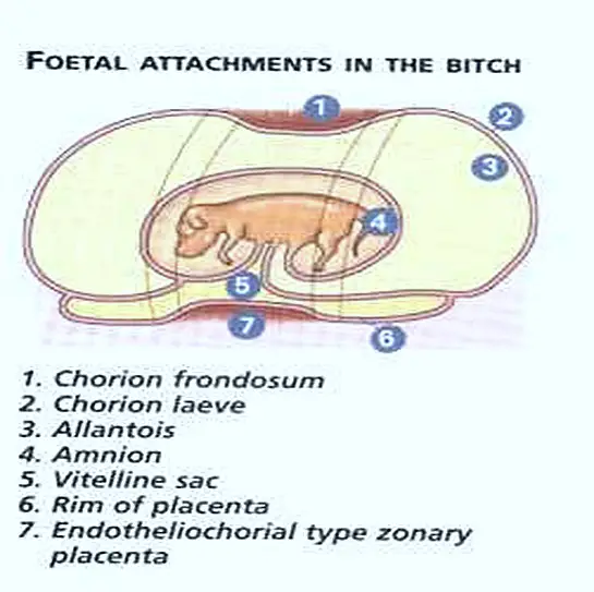
In these three types of placenta during perturition the foetus will not damage uterus. - Zonary placenta: Ex: Cat, Dog, Carnivores. The villi are In the form of transverse zones. In dog a single girdle of vhf will be present. In fox two girdles of villi are present. The villi penetrate Into uterine wall. Hence during parturitlon uterine wall Is damaged.
- Discoidal Placenta: Ex: Rat, Bat, Rabbit. On the entire surface of blastocyst the villi are in the form of discs. When the embryo Is growing It movesaway from uterus hence the with look like a disc. These villi are Intimately connected with uterus. Hence during parturitlon much uterine tissue is damaged.
CLASSIFICATION OF PLACENTA (BASING ON HISTOLOGY):
According to number of layers of cells present between foetus and uterus blood supply the placenta Is classified into five types.
a) Epithelio chorlal placenta: Ex: Pig, Horse, (Ungulates Lemmures)
The foetal chorion Is In contact with eplthelium of the uterus hence it is called epithello chorial placenta. In between foetal, maternal parts six layers are present.
- Endothelium of mother blood vessel.
- Maternal syndesmose connective tissue.
- Epitheliurn of mother
- Chorion of foetus.
- Foetus connective tissue (syndesmose
- Endothellum of foetal blood vessel.
If all the six layers are present the placenta is called epithello chorlal placenta.
b) Syndeumose chorial placenta: Ex: Sheep, Cow.
The allanto-chorianic vith will pierce into the uterus of the mother, the chorion will come in contact with syndesmose of mother’s uterus. Hence iti s called syndesmose chorial.
c) Endothelio chorial placenta: Ex: Dog, Carnivores. The chàrion of the foetus will come in contact with the endotheli of mother ‘s uterus, hence it is called endothelio-chorial placenta.
d) Hemochorial placenta: Ex: Bat, Man, Primates, Insectivores.
The placental connections are more lntimate. The chorion of foetus will float In the blood pools of mother’s utiras. Hence It Is called haemochorIal placenta.
e) Hemo endothelial placenta: Ex: Rat, Rabbit, Hence guinea-pig will float In mother’s blood. Hence it called hemo endothelial placenta.
Share this info graphic
<p><strong>Please include attribution to bioscience.pk with this graphic.</strong><br /><br /><a href='https://www.bioscience.pk/topics/zoology/item/732-placenta-in-mammals-structure-types-and-functions'><img src='https://www.bioscience.pk/images/Types.of.Placenta.in.Mammals.jpg' alt='placentainmammals' 540px border='0' /></a></p>
FUNCTIONS OF PLACENTA:
- Placenta will form a physiological barrier between mother and foetus. It will possess foetal and maternal blood mixing.
- Placenta allows the diffusion of monosacharides, amino adds, hormones, vitamins, oxygen, .carbondioxide, water and other waste materials, because of this it supplies food, oxygen to foetus.
- It works as an excretory organ of foetus. It releases the nitrogenous waste materials Into mother blood.
- It works as an endocrine gland. It will secretes lactogen ,progesterone,etc. hormones.
- The placenta will manufacture fructose from glucose.
Published in
Zoology
Tagged under
Monday, 03 July 2017 10:37
Teeth Modifications in Mammals

There is an interrelation between the form of teeth arid nature of food. These may be omnivorous, carnivorous, insectivorous herbivorous and rodents or gnawing. In Mammals dentition is complete in the first three types. Often incomplete in herbivores where the canines are absent. The absence of canines forms a gap between incisors and premolar. This is called diastema Rabbit). The canines are small or absent and the cheek teeth have broad grinding surfaces with transverse ridges - lophodont molars. In omnivorous mammals the incisors are suited for cutting the canines for tearing and cheek teeth with prominent crowns raised into rounded tubercles are adapted for crushing bunodont molars. In the carnivorous mammals, the incisors are either poorly developed or reduced and few in number and the canines are long and sharp suited for tearing and piercing the flesh But the molars with knife-like edges are modified for scratching the flesh from the bones. In the insectivores, the incisors of the upper and lower jaws become closely united to secure small active prey and the molars have sharp pointed crowns.
Depending upon the shape, size and number of cusps, the teeth are differentiated into:
- Monocuspici teeth - Teeth with one cusp or tubercie
- Bicuspid teeth - Teeth with two cusps.
- Tricuspid teeth - Teeth with three cusps and all the cusps are arranged in one plane by forming a triangle.
- Bunodont teeth - Teeth with a number of distinct blunt pointed cusps - Man and Monkey.
- Lophodont teeth - The cusps are fused in the form of ridges- Horse & Elephant.
- Selenodont teeth - Here the cusps are wear away, exposing the under lying dentineythich-getnuii by crescent -Shaped bands raf -
- Secodont teeth - The cusps are having sharp cuffing surfaces - carnivorous mammals. Carnassial teeth are present.
- Hypsodont teeth - The teeth are having long crown and deeply placed neck. - Horse
- Brachyodont teeth - Teeth with short crow, and neck at the surface gum. - Dog.
In sirenia (sea - Cow and Manatee), the teeth are devoid of enamel.
Published in
Zoology
Tagged under
Sunday, 02 July 2017 16:54
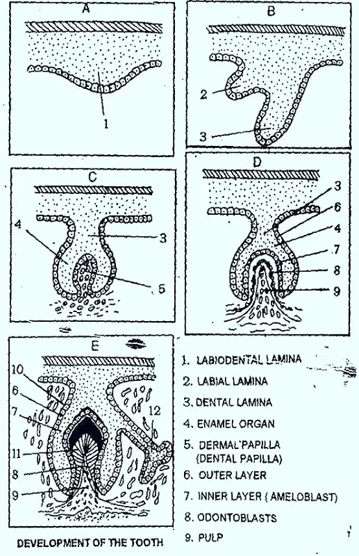
TOOTH DEVELOPMENT IN MAMMALS

The tooth develops partly from the epidermis and partly from the mesenchyme of dermis. Its enamel is derived from the epidermis and the remaining parts from the mesenchyme. Mammalian teeth develop in two sets. The milk or deciduous teeth develops from the buccal epithelium of the gums which are replaced later by the permanent teeth.The tooth develops partly from the epidermis and partly from the mesenchyme of dermis. Its enamel is derived from the epidermis and the remaining parts from the mesenchyme. Mammalian teeth develop in two sets. The milk or deciduous teeth develops from the buccal epithelium of the gums which are replaced later by the permanent teeth.
In the beginning the buccal epithelitim of the gum sinks down forming the t ridge. Simultaneously, the cells of malpighian layer of epidermis grow and move into the epidermis forming the dental lamina. Along the dental Lamina, the mesenchymal cells multiply and condense into a dental papilla or tooth bud. This grows upwards and pushes the dental kA Thereby an inverted cup form assumed The malpighian cells become markedly columnar and are known as ameloblasts. The mesenchymal ce are differentiated into a layer of odontoblast cells.
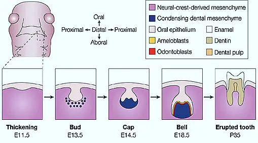
The tooth bud is now as ‘enamel organ’. The ameloblast cells secrete enamel’ towards their inner side, while the odontobiasts secrete ‘dentine’. The enamel sur rounds the upper part of dentine like a cap. The remaining mesenchyme cells of the dental papilla form the ‘pulp’ and the cavity of the enamel organ modifies into the pulp cavity. Sooner, the development of the tooth is completed,. The jaw bone forms a socket or alveolus around its base. The tooth increases in size by the secretion of more or mote dentine and finally emerges out from the gum. The odontoblasts now secrete cement around the neck and root of the tooth narrowing the pulp cavity. In majority of the mammals, the odontoblasts stop functioning after the development of tooth and the tooth stops growth. In some cases, like incisors; of rodents and tusks of elephants, the odontobiasts are functional throughout the life and the teeth continue to grow.
Published in
Zoology
Tagged under
Sunday, 02 July 2017 13:55
EVOLUTION OF MAMMALIAN MOLAR

Two main theories have been proposed to explain the evolution of molars.
- CONCRESCENCE THEORY OF ROSE
- TRLTUBERCULAR THEORY OF COPE & OSBORN
According to the concrescence theory, the molar tooth has evolved by the fusion of several simple cone-like teeth. This theory Is now abandoned in favor of the second with is now accepted with certain modifications.
Cope & Osborn theory is based on the paleontological evidence. As such the mammalian molar has been derived from a simple reptilian cone by the development of additional cusps. The Mesozoic mammal, the crown of the molar tooth has three cusps arranged more or less in the form of a triangle. This pattern of tooth has been termed tritubercular. It is assumed that the molars of the modem mammals have originated from the primitive tooth by the development of lateral cusps.
In the upper molars the three cusps are known as paracone, protocone, and metacone, The protocone lies on the apex at the inner side, the paracone lies external to it and the metacone to the back of the paracone. In the lower molars the cusps are named protoconid, paraconid and metaconid. The protoconid and metaconid are anterior and posterior to it in position. These cusps are joined by the ridges and later bell-like extensions or talons are produced from the ridges and additional cusps form on them. In this way, the various types of mammalian molars have evolved.
Published in
Zoology
Tagged under
Sunday, 02 July 2017 13:10
DENTITION IN MAMMALS

Teeth are the dermal derivatives of integument They are developed as a result of calcification in the mucous membrane of the 1 cavity. Along with the ridge of the two jaws, the teeth are arranged in a row. The teeth are present in almost all the mammals except in a few mammals In whale, the teeth are fused into plates and lost in the adult stage of Ant eaters. But in Echidna (spiny ant eater) the teeth are absent even in the embryo.
Structure of tooth: Each typical mammalian tooth is placed in the socket over the jaw bone. It is distinguished into three main pads.
- Root: It is the basal part embedded in the bony socket.
- Neck: it is the part above the root enclosed by the gum
- Crown It is the upper part beyond the surface of the gum.
The toot is separated from the socket by a vascular pridontal membrane. The vertical section tooth consists of the following parts.
- Pulp cavity: The entire tooth encloses a central pulp cavity surrounded by a layer of odontoblast cells filled with soft pulp. It is made up of connective tissue, blood vessels and nerve fibers.
- Dentin: A substance chemically similar to bone- dentine forms the major part of the tooth. But the dentine is permeated by numerous thin canaliculi.
- Enamel: It is present over the de in the crown and neck regions of the tooth. It is hardest and contains only traces of living matter.
- Cement: It surrounds the denting of the root portion of the tooth. It is bony in nature.
Published in
Zoology
Tagged under
Sunday, 02 July 2017 12:05
CHARACTERS OF MAMMALIAN DENTITION

The mammalian teeth are placed in the muscular sockets in the jaw bones. This type of dentition is called Thecodont. The teeth are ‘diphyodont’. Two sets of teeth develop during life lime. The first set of teeth develop in the young and known as ‘milk or deciduous teeth’, after certain age these are replaced by the second set of permanent teeth. But in bats ‘and, guinea pigs the milk teeth are shed before birth. In platypus, toothed whales, sloths and sirenians the monophyodont dentition’ is present. Milk dentition is reduced by the replacement of the third premolar in marsupials.
All the teeth are not alike in all mammals. Mostly mammals exhibit ‘heterodont dention’. The teeth are modified in the form and function. But in Dolphins and porpoises it is homodont (all teeth are alike). There are four types of mammalian teeth.
1) Incisors: These are present on the premaxillae bones of the upper jaw and dentary bones of the lower jaw. They have only one ro and sharp cutting edges. So this type of teeth is used in sizing and cuffing.
2) Canine: These are present between the incisors and premolar. There is a single such troth in each half of each Jaw. These are long, conical teeth with a single root and simple, sharp-pointed crown.
3) Premolars : These follow the canines. They have double root and compressed crown with one or two cusps. These possess grinding and crushing surfaces. Premolars are replaced once in life time.
4) Molars: These are next to the premolars. They have more than two roots and cusps. These do not have predessors and always develop in the permanent set. Molars are used for crushing and mastica lion.
Both prermolars and molars are collectively knows, as cheek teeth and are borne on maxillae and dentaries. These have broad crushing and grinding surfaces or cusps.
Published in
Zoology
Tagged under
Sunday, 02 July 2017 11:37
Dental Formula in Mammals

Mostly the number of teeth is fixed in each mammalian species. Mammalian heterodont dentition is expressed by a "dental formula". The number and arrangement of teeth in each half of the upper and lower jaws are constant and identical. Hence the teeth can be expressed by using the initials - I, C, Pm, and P4. The number of teeth differs in the various orders of mammals and is closely related to their feeding habits.
Published in
Zoology
Tagged under
Sunday, 02 July 2017 10:39
SKIN OF MAMMALS (RABBIT)

SKIN STRUCTURE AND FUNCTIONS IN MAMMALS
The body of Rabbit is covered by skin. It shows two layers,
- Epidermis
- Dermis
Associated with skin are hair, claw and glands.
1. Epidermis: It is the upper part of the skin. It is divisible into two parts
a) Stratum comeum: This layer is hard scale like, dead keratinized and flat.
Keratin prevents the passage of water and solutes. This layer is cast off periodically. Below this layer transparent layer is present It is called stratum lucidum. In this layer - iucidin substance is seen When stratum comeum is removed this lucidum layer will become stratum comeum Below the stratum lucidum another layer is present. It is called stratum granulosum. It contains granular cells. Final layer of epidermis is stratum germinatum. It shows columnar cells. These cells can undergo division.
In the epidermis special pigment cells are present they are called chromatophore cells.
2. Dermis: It is present below the epidermis It is tough and flexible. It is made by connective tissue, unstrapped muscles blood capillaries, nerves and fat cells This part of the skin is in contact with nerve endings.
Glands: In the skin of Rabbit sweat or sudorif glands and sebaceous glands are present.
The sebaceous glands are sac like glands. They are branched. They secrete oily secretion which keeps the hair and skin water proof.
The sweat glands are thin and long tubes. They arc coiled the sweat glands are found deep in the dermis. They open on the surface of the skin. These glands will secrete salty, watery solution from blood. It is excretory in function. This gland plays an important role in the regulation of body temperature.
Hair: It is epidermal in origin. It is seen only in mammals. It develops from the stratum germinatum. It has two parts root and shaft the hair is enclosed in a tube like hair follicle. At the base of the follicle dermal papilla is present. It is supplied with blood vessels.
The skin covers the body and protects the body organs
Skin Functions:
- It covers the body parts.
- Sweat glands are excretory and regulate the body temperature.
- Skin works as tactile sense organ.
- Sebaceous glands are modified into mammary glands which secrete milk for young ones.
Published in
Zoology
Sunday, 02 July 2017 10:31
STERNUM OF RABBIT

In the mid ventral region of thorax of Rabbit sternum is present. It is made by five bony pieces called "Sternabrae". It is called "mesosternum". This will show "presternum" anteriorly. It is a long bony piece. It is connected with clavicles. On the lower side mesosternum it articulates with xiphi-sternum, which ends with xiphoid cartilage.
The ribs articulated with sternum by their cartilage part. In rabbit the first pair of ribs are attached to the presternum.
Uses of sternum :Thus sternum gives strength and support to the body on the ventral side. It helps in the process of respiration also.
Published in
Zoology
Tagged under
Sunday, 02 July 2017 03:08
ORGAN OF CORTI OR COCHLEA

The internal ear of rabbit includes "organ of corti" or Cochlea spirally coiled. Lagena is enclosed and fused with the spirally coiled j the preotic bone which form cochlea. Cochlea is a bony tube line connective tissue. The cavity of the cochlea is transverse by two brines. It is divided into 3 cavities. They are
- Scala vestibuli
- Media
- Scala tympani.
The middle cavity is filled with endo lymph membranes that cover the Cochlea are Reisner's membrane and membrane. The epithelium of basilar membrane is highly modified. It is "organ of corti". It has hair cells and supporting cells. It is supported by cochlear branch of 8th cranial nerve. It is useful for hearing.
Published in
Zoology
Tagged under
Saturday, 01 July 2017 20:19
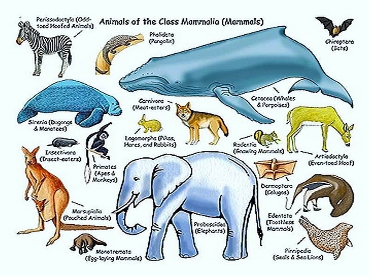
CLASSIFICATION OF MAMMALS: THERIA
THERIA
This group includes Mammals which show true mammals characters
Infraclass Metatheria:
They show almost all the Mammalian characters.
- Females show brood pouch or Marsupium on their ventral abdomen.
- Sebaceous glands are modified into mammary glands.
- Pelvic girdle shows epipubic bones. This supports Marsupium.
- Ribs are two headed.
- Carpus callosum is absent.
- Vagina and uterus are double (didefphic condition)
- They give birth to immature young ones.
- This infra class includes 1 order.
Order Marsupialia:
Ex: a) Didelphis – Opossum, b) Dasyurus - Tiger cat, c) Paramales - Bandicoot, d) Macropus - Kangaroo, e) Notorgetes - Marsupial mole
Infraclass Eutheria:
- Mammary glands are well developed.
- Double headed ribs are seen.
- Carpus callosum is present.
- Urinogenital organs will open independently into the Rectum.
- Testis are extra abdominal.
- Uterus and vagina are single.
- They show placenta in the development of embryo. This infra class is divided into following orders.
1. Order: Insectivora:
- Small animals are seen in this order.
- Snout is long.
- Five toed feet are seen.
- They are Nocturnal and Terrestrial
Ex: 1) Paraechinus (Hedgehog), 2) Talpa (mole), 3) Echinosorex (Shrews)
2. Order: Dermopetera:
- In between fore limbs and hind limbs, in between head and fore limb, in between hind limbs and tail, membrane is formed. This helps for gliding in air.
- They are Nocturnal.
Ex: Galeopithecus (Flying lemur)
3. Order: Chiroptera:
- Fore limbs are modified wings.
- Hind limbs are weak.
- Digits are clawed.
- Eyes are small and vision is very weak.
- They are usually called Bats.
Ex: Pteropus (Flying fox), Megaderma (Vampire bat)
4. Order: Primates:
- Hands and feet are more or less prehensile.
- Thumb is oppossable to other digits.
- Only two pectoral mammary glands are present.
- Cerebrum is big and convoluted.
Ex: 1) Chyromys (Aye aye), 2) Loris, 3) Hylobates (Gibbon), 4) Anthropithecus (Chimpanzee), 5) Simia satyrus (Orangutan), 6) Macaca (Monkey), 7) Homo Sapiens (man)
5. Order: Edentata:
- Feet has 5 digits. They are clawed.
- Testis are abdominal.
Ex: 1) Dasypus = ormadilla, 2) Bradypus = Three toed sloth, 3) Myrmecophaga' = Giant ant eater
6. Order: Pholidota:
- The body is covered by scales.
- In between scales hairs are present.
- Teeth are completely absent.
- Tongue is long, sticky and protrusible (Helpful to catch the ants).
Ex: 1) Manis (scaly ant eater or parigolin)
7. Order: Lagomorpha:
- Small mammals
- Tail is short.
Ex: Oryctologus - Rabbit, Lepus (Hare)
8. Order: Rodentia:
- Small mammals.
- Canines are absent. (Diastema is present)
Ex: Ratus ratus - Rat, Funambulus - Squirrel, Hystrix - Porcupine.
9. Order: Cetecea:
- Large aquatic animals.
- Body is spindle shaped and fish like.
- Hair is absent.
- Neck is absent.
- Fore limbs are modified into paddles.
- Hind limbs are absent.
Ex: Orcinus - Killer whale, Physeter - Sperm whale (Largest toothed whale) Plantanista – Dolphin
10. Order: Sirenia:
- These are all aquatic mammals. They are called sea - cows.
- Hind limbs are absent.
- Testis are abdominal.
Ex: Halicore dugong (dugong- sea cow), Trichecus (Manatee)
11. Order: Carnivora:
- They are terrestrial, arborial or aquatic mammals.
- Mammary glands are present on abdomen.
- The last premolar is called carnassial teeth.
Ex: 1) Panthera leo - Lion, 2) Pathera tigris - Tiger, 3) Acinomyx - Leopard, 4) Vulpes - Fox, 5) Trichecus - Walrus.
12. Order: Tubulidentate:
- Body is stout and pig like.
- Skin is covered by thick and less hair.
- Ears are long, errect and pointed.
Ex: Orycteropus
13. Order: Proboecide:
- Largest Terrestrial Mammals.
- Long probosis is seen.
- Incissors will become Tusks.
Ex: 1) Elephas - Indian elephant
14. Order: Hyrocoidea:
- Small Rabbit like
- Ears arc short
Ex: Procavis
15. Order: Perissodactyla:
- Single Looted mammal.
- The middle light of limbs will develop hoof.
Ex: 1) Equas (Horse), 2) Rhinoceros – Rhinoceros
16. Order: Artiodactyla:
16. Order: Artiodactyla:
- Terrestrial, semi aquatic animal.
- Limbs have two hooves.
Ex: Hippopotamus - Horse of River Camelus - Camel, Sus - Pig
Published in
Zoology
Tagged under
Saturday, 01 July 2017 20:06
CLASSIFICATION OF MAMMALS: PROTOTHERIA

Mammalia is divided into two subclasses. 1) Prototheria 2) Theria
GENERAL CHARECTERS OF PROTHERIA
- External pinnae are absent.
- In the young stage teeth are present and in the adults horny plates are formed.
- Mammary glands are present and tears are absent.
- Body is covered by hairs.
- Pelvic girdle shows epipubic bones.
- Vertebrae will not show epiphyses.
- Ribs are single headed. (Tuberculum is absent)
- Corpus callosum is absent in brain.
- Cochlea is simple and is not coiled.
- In the males the testis is abdominal.
- Females are egg laying. Hence prototherians are called egg laying Mammals.
This subclass includes only one order that is Monotremata.
Order Monotremata:
Thesemammals show prototherian characters. They can found in Australia, Tasmania and New-guinea.
Ex: 1. Echidna (Spiny ant eater), 2. Ornithorhyncus (Duck billed platypus)
Published in
Zoology