Venous System of Rabbit
Explore the intricate rabbit circulatory system with our visual diagram. Understand the vital role of valves in maintaining proper circulation.
Rabbits, those adorable creatures known for their twitching noses and remarkable agility, harbor intricate systems beneath their furry exteriors. One such system, the venous system, plays a pivotal role in maintaining their vitality. In this article, we embark on a journey to uncover the mysteries of the venous system of rabbits. Delving into the circulatory intricacies, we'll navigate the blood-vascular pathways and shed light on how this system operates.
Exploring the Circulatory System of Rabbits
The circulatory system, also referred to as the cardiovascular system, serves as a transportation network within a rabbit's body. Comprising the heart, blood vessels, and blood itself, this system ensures the efficient delivery of oxygen, nutrients, and waste removal. At its core lies the venous system, responsible for returning deoxygenated blood to the heart for oxygenation.
Unveiling the Blood Vascular System of Rabbits
At the heart of the circulatory system are three types of blood vessels: arteries, veins, and capillaries. Arteries act as expressways, carrying oxygenated blood away from the heart to various tissues. Veins, on the other hand, form the return route, ushering deoxygenated blood back to the heart. Connecting these vessels are capillaries, intricate networks that facilitate nutrient and gas exchange.
Deep Dive into the Venous System of Rabbits
Veins, often deemed the unsung heroes of circulation, possess unique features. Structurally, they boast thinner walls and larger lumens compared to arteries. The journey of deoxygenated blood through the venous system is a meticulous process orchestrated by both physiological and anatomical elements.
The venous system operates through a series of interconnected pathways, each serving as a conduit for blood transport. Gravity and muscular contractions aid blood movement, but a remarkable component takes center stage: valves. These valves, akin to miniature gates, prevent backward blood flow, ensuring blood reaches its intended destination.
The Journey of Blood: Circulation Process in Rabbits
The circulation process commences with oxygen-rich blood being pumped out of the heart through the aorta, the largest artery. As arteries branch out, they progressively narrow into arterioles and capillaries, allowing the exchange of nutrients and oxygen with surrounding tissues. Subsequently, deoxygenated blood enters venules and veins, embarking on its return expedition to the heart.
Adapting to Unique Rabbit Physiology
Rabbits boast a unique physiology that sets them apart from humans. Their heart possesses four chambers, much like ours, but their circulatory demands differ. Rabbits exhibit a lower blood pressure than humans, a feature attributed to their herbivorous diet and size. This distinct physiology influences their venous system's architecture, reflecting evolutionary adaptations.
Rabbit blood vessels, especially veins, endure fluctuations in pressure due to their agile movements and diverse postures. This variance demands robust venous walls and efficient valves to counteract the challenges posed by gravity and rapid motions.
The blood from various parts of the body is collected by different types of veins which constitute the venous system. Coronary veins collect the venous blood from the wall of the heart into the left precaval vein.
The blood from the anterior and posterior ends of the body is mainly collected by two precavals or superior vena cavae and one post caval or inferior vena cava respectively.
Each precaval is formed by the union of three veins namely;
- External jugular vein
- Internal jugular vein
- Subclavian vein
The external jugular vein brings the blood from the tongue, jaws, and muscles of the head.
- The external jugular is connected with its fellow on the opposite side by a transverse or jugular anastomosis that anastomoses in the neck.
- The internal jugular vein receives the internal carotid vein collecting blood from the brain and neck.
- Then it runs down the neck along the side of the trachea and opens into the external jugular vein opposite to the subclavian.
- The subclavian vein is a large vein on each side of the body. It collects blood from the shoulders and forelimbs.
- The two precavals open into the right auricle.
Each precaval vein also receives two smaller veins on either side namely; (a) Anterior intercostal vein and (b) Azygos cardinal vein.
- The anterior intercostal vein collects blood from the anterior intercostal spaces and joins the precaval.
- This vein is formed by the union of four or five smaller veins.
- Azygos cardinal vein is formed by the union of small veins and opens into the right precaval vein.
- On the left side of the animal, the Azygos vein is absent, but a few small veins form a hemizygos vein.
- The Hemizygos vein joins the right azygos vein through a transverse Anastomosis.
- The post-caval vein or Inferior vena cava is a large median vein formed in the posterior part of the abdominal cavity by the union of several veins.
- The internal iliac or hypogastric of the two sides unite along the middle line together with a median caudal vein from the tail to form a common hypogastric vein.
- The internal iliac brings the blood from the back of the thighs.
- The common hypogastric vein receives a large external iliac vein.
on either side. Each external iliac vein is formed by a femoral vein bringing blood from the inner or preaxial side of the thigh and a posterior epigastric vein collecting blood from the ventral wall of the abdomen.
- A small vesicular vein also brings the blood from the urinary bladder into the common hypogastric vein.
- The post-caval vein runs anteriorly and it receives several veins from different organs on its way to the right auricle.
- In front of the external iliac, the post-caval vein receives a pair of large ilio-lumbar veins which bring blood from the hind part of the walls of the abdomen.
- Anterior to the iliolumbar a pair of gonadial veins also open into the post caval, spermatic veins from the testes of males, and ovarian veins from the ovaries of females.
- In front of the gonadial veins, the post caval receives two large renal veins collecting blood from the two supra renal and kidneys.
- The right renal vein is slightly anterior than the left renal vein.
- The renal portal system is absent in the rabbit. Post caval vein further runs anteriorly through the liver from which it receives blood from several hepatic veins.
- The hepatic portal system is present in rabbits and it consists of a hepatic portal vein ventral to the post caval vein.
- It enters into liver and divides into several branches to supply to various lobes of the liver.
The hepatic portal vein is formed by several veins like (i) a lienogastric vein that brings blood from the stomach and spleen, (ii) a duodenal vein collecting blood from the duodenum and pancreas, (iii) an anterior mesenteric vein bringing blood from the caecum, colon and part of the rectum, and (iv) posterior mesenteric vein that collects blood from lower part of the rectum.
- The blood from the lungs is collected by a pair of large pulmonary veins.
- The two pulmonary veins open by a common aperture into the left auricle.
- The pulmonary veins bring oxygenated blood from the lungs into the left auricle.
Visualizing the Circulatory System: Rabbit Diagram
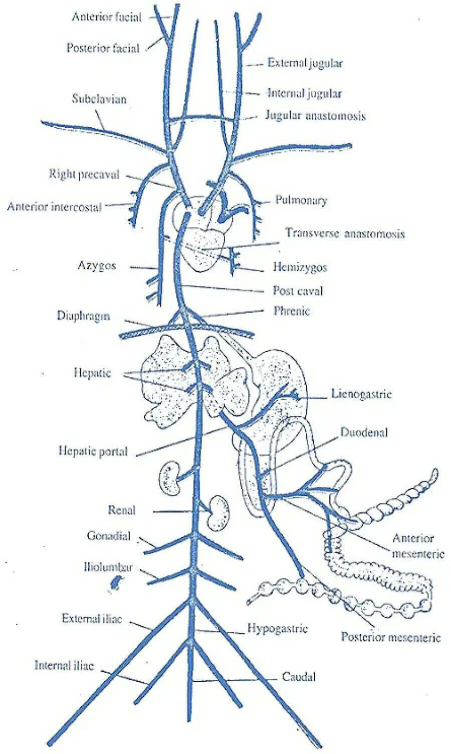
The above diagram (Figure 241.1) illustrates the intricate pathways of the circulatory system within a rabbit's body. The diagram is a comprehensive visual representation that showcases the journey of blood as it flows through various blood vessels, highlighting the key components and connections of the rabbit's circulatory system.
In the diagram, arteries are depicted as branching out from the heart, carrying oxygenated blood to different parts of the rabbit's body. These arteries gradually narrow into smaller arterioles and then further into capillaries, forming a network that facilitates the exchange of nutrients, gases, and waste products with surrounding tissues.
As the diagram unfolds, deoxygenated blood is seen making its way back to the heart through venules, which merge into larger veins. The veins are prominently illustrated, and special attention is given to the presence of valves within them. These valves play a crucial role in preventing the backward flow of blood, ensuring its steady movement towards the heart even in the face of challenges posed by gravity and the rabbit's movements.
The above diagram (Figure 241.1) serves as an educational tool, allowing viewers to gain a deeper understanding of how the rabbit's circulatory system operates. The strategic arrangement of arteries, veins, capillaries, and valves in the diagram provides a clear visual guide that aids in comprehending the complexities of blood circulation in rabbits.
Conclusion
In our exploration of the venous system of rabbits, we've embarked on a journey through the intricate circulatory pathways that sustain these furry creatures. From arteries delivering vitality to veins returning blood laden with waste, every element plays a vital role. The rabbit's unique physiology and evolutionary adaptations further underscore the marvel of its venous system.
The information on this page is peer reviewed by a qualified editorial review board member. Learn more about us and our editorial process.
Last reviewed on .
Article history
- Latest version
Cite this page:
- Comment
- Posted by Dayyal Dungrela