Life History of Fasciola Hepatica (Sheep Liver Fluke)
Fasciola Hepatica is digenetic parasite. It completes its life history in sheep and freshwater snails. Sheep is the primary host and snail acts as an intermediate host. Johan de Brie" in the 18th century identified Fasciola in the sheep liver. Leuckart-Thomos reported the life cycle of liver fluke in snails.
The systematic position of Fasciola:
- PHYLUM: PLATYHELMINTHES
- CLASS: TREMATODA
- ORDER: DIGENEA
It is distributed throughout the world. It attacks sheep and causes liver rot disease in them.
The life cycle of Fasciola hepatica is completed within two hosts. The primary host, is the sheep, while the intermediate host, is a snail. This type of life cycle, involving two hosts, is termed a digenetic parasite.
The liver fluke undergoes copulation in the bile duct of sheep. During copulation, the penis of one liver fluke is inserted into the laborer's canal of another fluke and sperms are transferred. In the oviduct fertilization takes place. In liver fluke cross fertilization takes place by copulation. Self-fertilization also takes place.
The fertilized egg is surrounded by yolk cells and developed into shelled eggs or capsules. The shelled egg is light brown. It shows a lid. It is 130 to 150 microns in length. These eggs will reach the intestine through the bile duct. They will be sent out of the body of sheep through fecal matter. Thousands of eggs are liberated by sheep every day. These eggs will undergo early development when the environmental conditions are favorable. At 25 degrees centigrade, the egg gives rise to a Miracidium larva in 9 to 15 days.
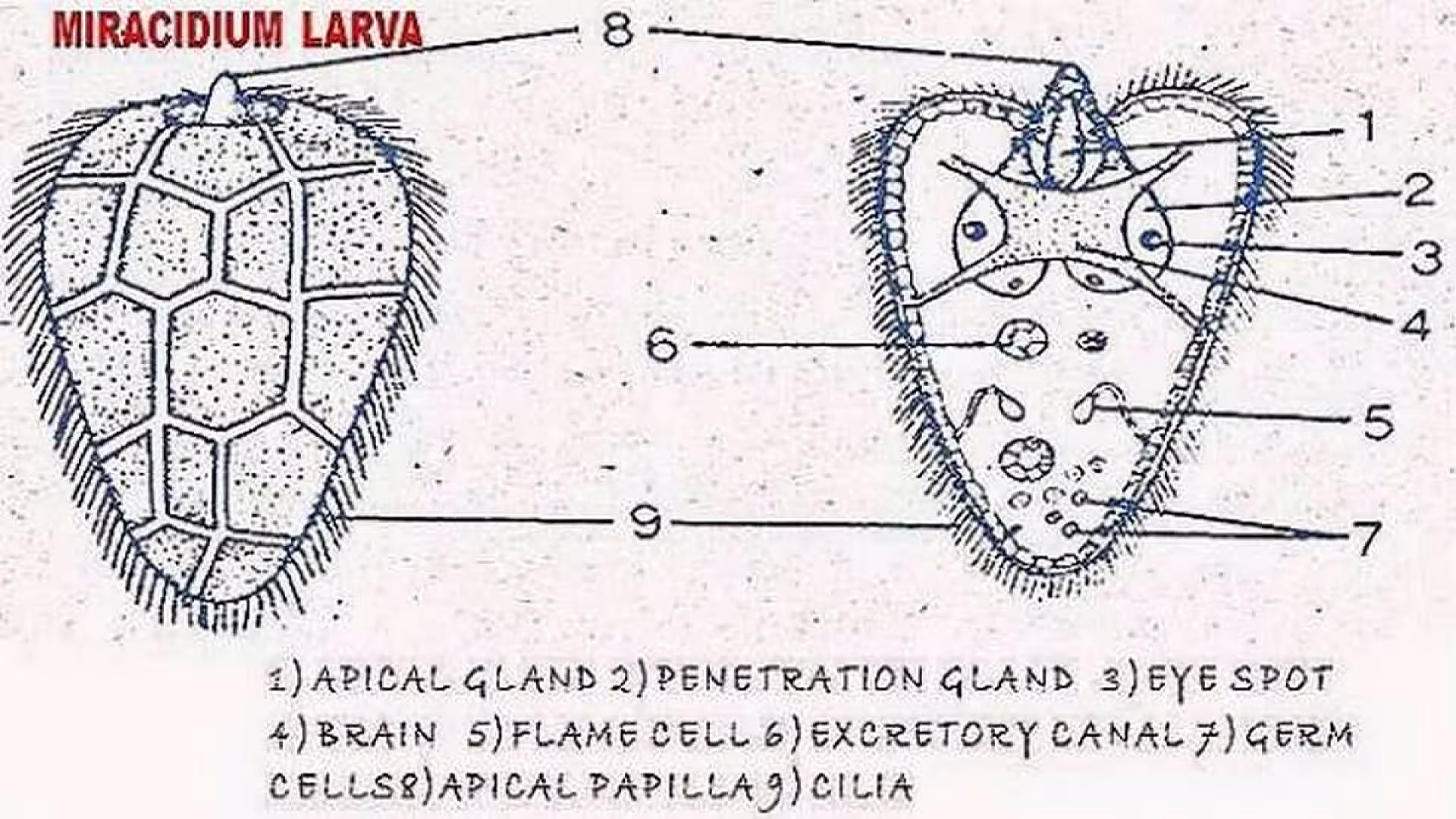
MIRACIDIUM LARVA:
In the life history of a liver fluke, the first larval stage is miracidium when the egg is placed in water the lid opens and miracidium comes out. This larva swims in the water.
Characters:
- It is 150 microns in length. It is small. It is conical in shape.
- It is covered by ciliated epidermal cells.
- The body is covered by 21 ciliated cells which are arranged in five rows.
- First row - six cells
- Second row - six cells
- Third row - three cells
- Fourth row - four cells
- Fifth row - two cells
- With the help of cilia it swims in the water.
- At the apex of the larva an apical papilla or boring papilla is present.
- An apical gland in the miracidium larva opens into the apical papilla. On either side of it, two penetration glands are present.
- A brain is present. Above the brain two eye spots are present.
- A pair of flame cells are present which open out laterally towards the posterior end. The larva shows several germ cells.
- The miracidium larva lives only for 8 hours. During this time it will swim in search of the secondary host.
Transmission to secondary host:
The secondary host of liver fluke is Lymnaea truncatula or Planorbis (freshwater snails. When the miracidium larva comes in contact with the snail it pierces into the soft body of the snail. Apical papilla and secretions of the penetration gland will help the larva to bore into the body of the snail. In the body of a snail, miracidium develops into the sporocyst stage.
Sporocyst:
In the body of a snail, miracidium enters into the pulmonary sac. Their miracidium will lose its ciliated epidermis. It becomes a bag-like structure. It loses all the structures except flame cells and germ cells. The germ cells will undergo parthenogenesis and give rise to the next larvae called Redia. The sporocyst absorbs nourishment from the host tissues and often destroys the host.
Redia:
In the sporocyst, five to eight redia larvae are produced. They come out of the sporocyst by rupturing the wall of the sporocyst. This larva is elongated in structure. It is covered by a thin cuticle. It shows a muscular collar. It helps in locomotion. Near the collar a birth pore is present. The next larval stage will go out through the birth pore. The larva shows a gut that opens out through the mouth. The mouth opens into the pharynx which leads into the intestine. Many flame cells are present. The flame cells of one side will open into a common excretory duct which opens out through a single nephridiopore. The mesenchyme of the larva shows germ cells.
The germ cells will undergo parthenogenesis and give rise to the next larval stage called cercaria in the winter season. These cercaria larvae will come out of the redia through the birth pore.
Cercaria:
The redia larva will give 15 to 20 cercaria larvae. They are liberated from the redia larva through the birth pore.
- It is oval with a tail.
- It is 0.25mm to 0.35mm in length.
- The cuticle covering will show backwardly directed spines.
- Two suckers are present, a) Oral sucker around the mouth, b) ventral sucker.
- The digestive system starts with the mouth and opens into the pharynx, esophagus, and intestine. The intestine is divided into two branches.
- More flame cells are present. All of them open into excretory tubules. The two excretory tubules will unite at the posterior end and become excretory bladder. It gives an excretory tube. It divides into two, which open out through the nephridiopore.
- Germ cells are present.
The completely developed cercariae will enter into water from the body of the snail. They swim for 2 or 3 days in the water and settle on a water plant.
Metacercaria:
Cercaria larva after attaching to a water plant loses its tail and develops a cyst around itself. It is called Metacercaria. It is 0.2 mm in diameter. These stages can develop only when they enter into sheep. This stage can survive for a few weeks if they are present near water.
Transmission:
When the sheep eats the plants with metacercaria stages they enter into its digestive system. The cyst wall is digested in the intestine, it penetrates through the intestine wall and reaches the liver. It takes six weeks to grow into an adult. It takes 12 weeks to attain sexual maturity.
Thus Fasciola hepatica completes its life cycle.
Pathogenesis:
- It causes hepatitis in sheep.
- It causes hemorrhage in sheep.
- It causes liver rot disease in sheep. Because of this, the sheep become weak.
The information on this page is peer reviewed by a qualified editorial review board member. Learn more about us and our editorial process.
Last reviewed on .
Article history
- Latest version
Cite this page:
- Comment
- Posted by Dayyal Dungrela