It is a type of internal bud. Gernmules are formed in fresh water sponges. Eg: Spongilla. During unfavourable conditions the archaeocytes of the body (some people say 'trophocyte' of the body) will group together. These archaeocytes are filled with glycoprotien or lipoprotien. Around this group protective layers are formed.
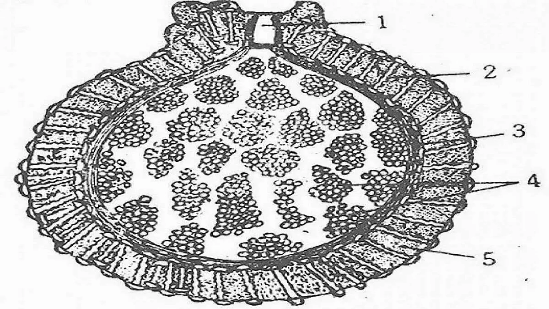
Each gemmule shows a central group of archaeocytes. Around which an inner and outer chitinous layers are present. Around this a pneumatic coat is formed. This coat is supported by small Amphidise speckles. The outer coat shows an opening called "Micropyle".
When a number of gemmules are formed, the animal disintegrates and all the gemmules are liberated out. They lie in dormant condition on the bottom of the pond.
With the advent of favorable conditions the gemmules hatch and develop into new sponges.
The information on this page is peer reviewed by a qualified editorial review board member. Learn more about us and our editorial process.
Last reviewed on .
Article history
- Latest version
Cite this page:
- Comment
- Posted by Dayyal Dungrela