Bleeding Time (BT) and Clotting Time (CT)
BT CT test (bleeding/clotting time test) measures bleeding and clotting duration. Learn procedures, normal ranges, and how these tests detect blood disorders.
Highlights
- What You’ll Read: A detailed analysis of the BT and CT test, bleeding time procedures (including Duke method), and clotting time protocols.
- What You’ll Learn: BT CT test procedure, results, normal ranges, and diagnosis platelet or clotting disorders.
- What You’ll Discover: The clinical role of BT CT test in detecting bleeding issues and why Duke method is outdated.
The BT CT test refers to two distinct diagnostic procedures that collectively evaluate the hemostatic system. The BT test quantifies platelet-mediated hemostasis by measuring the duration required for small blood vessels and platelets to halt bleeding following a standardized incision. In contrast, the CT test assesses the efficiency of the coagulation cascade by determining the interval between vascular injury and fibrin clot formation. Together, they evaluate blood’s ability to prevent excessive bleeding.
The Bleeding Time test specifically evaluates platelet functionality and the initial phases of hemostasis, which are critical for preventing excessive hemorrhage. Clinically, it serves as a diagnostic tool for identifying disorders such as thrombocytopenia (abnormally low platelet counts) and von Willebrand disease (a deficiency in platelet-adhesive glycoproteins). Additionally, it is utilized to monitor therapeutic interventions targeting platelet activity. This test is particularly sensitive to disruptions in primary hemostasis, making it indispensable in the evaluation of unexplained bleeding tendencies.
Conversely, the Clotting Time test focuses on secondary hemostasis by analyzing the integrity of coagulation pathways, including intrinsic and extrinsic mechanisms. It is pivotal for diagnosing coagulation disorders such as hemophilia (factor VIII or IX deficiencies) and for evaluating the efficacy of anticoagulant therapies (e.g., heparin or warfarin). By measuring the time required for fibrin clot formation, the CT test reflects the functional status of clotting factors and enzymatic cascades. When interpreted alongside BT results, it facilitates a comprehensive assessment of hemostatic balance, encompassing both cellular (platelet-driven) and plasma-based coagulation processes.
1. BT Test (Bleeding Time)
The BT test is conducted using one of the following methods:
- Duke’s Method
- Ivy’s Method
- Template Method
Among these, the Template method provides more reliable results compared to the Duke’s method, which is no longer widely used due to the risk of hematoma formation and lack of standardization. However, the Ivy’s method remains commonly used, especially in clinical laboratories.
1. Duke’s Method for Bleeding Time
Principle
The bleeding time Duke’s method measures platelet function by timing bleeding cessation after an earlobe or fingertip puncture.
Required Equipment
Sterile lancet, filter paper, stopwatch, antiseptic (e.g., alcohol), and gauze for wound care.
Procedure
- Clean the earlobe/fingertip with alcohol.
- Make a 3–4 mm deep puncture using a lancet.
- Blot blood every 15–30 seconds without touching the wound.
- Record time until bleeding stops (normal range: 1–3 minutes).
Limitations: Inconsistent due to variable puncture depth; largely replaced by Ivy’s method for standardized results.
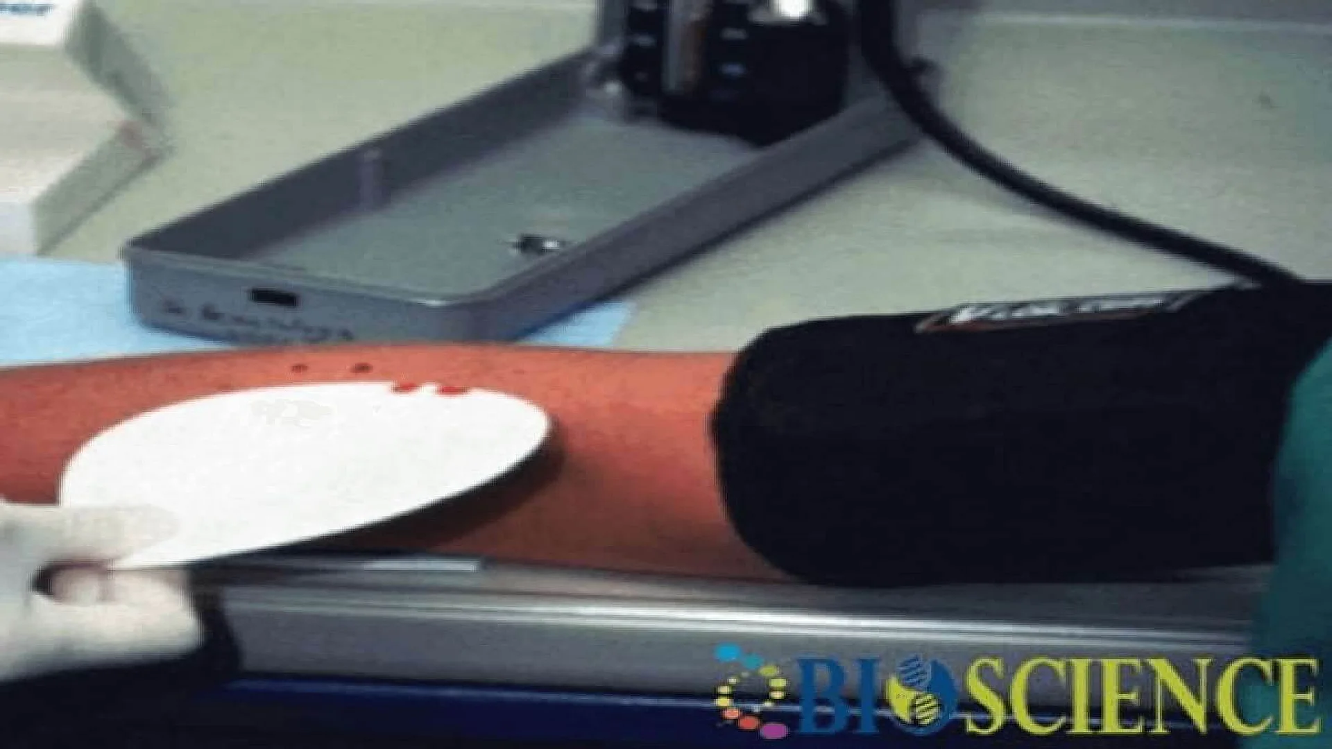
2. Ivy’s Method for Bleeding Time
Principle
In Ivy’s method, three punctures are made on the volar (inner) surface of the forearm using a lancet. The time it takes for bleeding to stop from these punctures is measured. The pressure is maintained between 30-40 mm Hg using a sphygmomanometer to ensure standardized conditions.
Required Equipment
- Sterile disposable lancets
- Sphygmomanometer
- Filter paper
- Stopwatch
Procedure
- Measure the patient’s blood pressure to ensure it is within the normal range.
- Clean the volar surface of the forearm with 70% ethanol and allow it to dry.
- Make three punctures, spaced about 5 cm apart, avoiding superficial veins and scars.
- Start the stopwatch immediately after the first puncture is made.
- Absorb the blood with filter paper at 15-second intervals, without touching the wound.
- Stop the timer when blood no longer stains the filter paper.
- Record the average time from all three punctures and report it as the bleeding time.
3. Template Method for Bleeding Time
Principle
In Template method a surgical blade and disposable template is used to create a standardized incision depth and length, ensuring consistent measurement of platelet plug formation.
Required Equipment
- Surgical template (e.g., 5–10 mm slit for uniform incision).
- Sterile surgical blade or automated incision device.
- Sphygmomanometer (to maintain 40 mmHg pressure).
- Stopwatch, filter paper, antiseptic (70% ethanol).
Procedure
- Apply a blood pressure cuff to the upper arm (40 mmHg) to standardize venous pressure.
- Clean the volar forearm with ethanol and let it dry.
- Place the surgical template on the skin and make a 1 mm deep incision using the blade.
- Start the stopwatch immediately and blot blood with filter paper every 15 seconds.
- Record the time when bleeding stops (normal range: 2–7 minutes).
Advantages:
- More reliable than Duke’s method due to standardized incision depth/length.
- Reduces variability in results, improving diagnostic accuracy for platelet disorders.
Normal Bleeding Time Range
- 2 to 7 minutes.
- Most individuals will have a bleeding time of less than 4 minutes. If bleeding persists for more than 20 minutes, the test is stopped, and the result is reported as >20 minutes.
Prolonged Bleeding Time
Prolonged bleeding times may indicate various medical conditions, including:
- Thrombocytopenia (low platelet count)
- Von Willebrand Disease
- Platelet dysfunction disorders
- Blood vessel disorders
- Afibrinogenemia (absence of fibrinogen)
2. CT Test (Clotting Time)
- Blood is drawn into a tube and observed at 37°C until clot formation.
- Normal CT Range: 8–15 minutes.
BT CT Normal Ranges
| Test | Gender | Age-Group | Normal Range | Prolonged Result Indicates |
|---|---|---|---|---|
| BT Test | Unisex | All age groups | 2–7 minutes | Thrombocytopenia, platelet dysfunction |
| CT Test | Unisex | All age groups | 8–15 minutes | Hemophilia, vitamin K deficiency |
Takeaways
- BT test focuses on platelet function; CT test assesses clotting factors.
- CT test is usually performed to diagnose conditions like hemophilia or disseminated intravascular coagulation (DIC).
- Abnormal BT CT test results guide further testing (e.g., factor assays, platelet count).
FAQs
What is the BT test?
What is the CT test?
How is the BT test performed?
What is the normal range for Bleeding Time (BT)?
What is the normal Clotting Time (CT)?
What conditions can extend the Bleeding Time (BT)?
What are the common methods to measure Bleeding Time (BT)?
What factors can affect Clotting Time (CT)?
Why is the BT test not recommended if the platelet count is low?
What are the differences between Duke’s, Ivy’s, and Template methods for Bleeding Time?
The information on this page is peer reviewed by a qualified editorial review board member. Learn more about us and our editorial process.
Last reviewed on .
Article history
- Next review due:
- Latest version
Reference(s)
- BL, Evatt., et al. “Fundamental Diagnostic Hematology: The Bleeding and Clotting Disorders.” 2nd ed., US Department of Health and Human Services, 1992
- SM, Lewis., et al. “Dacie and Lewis’s Practical Haematology.” 9th ed., Churchill Livingstone, 25 October 2001, isbn: 9780443063770.
Cite this page:
- Posted by Dayyal Dungrela