Hormone Waves Reshape the Brain: MRI Study Shows White and Gray Matter Shift Across the Menstrual Cycle
New research reveals that normal menstrual-cycle hormone fluctuations are linked to rapid, measurable changes in white and gray matter across the entire brain, highlighting the brain’s dynamic response to endocrine rhythms.
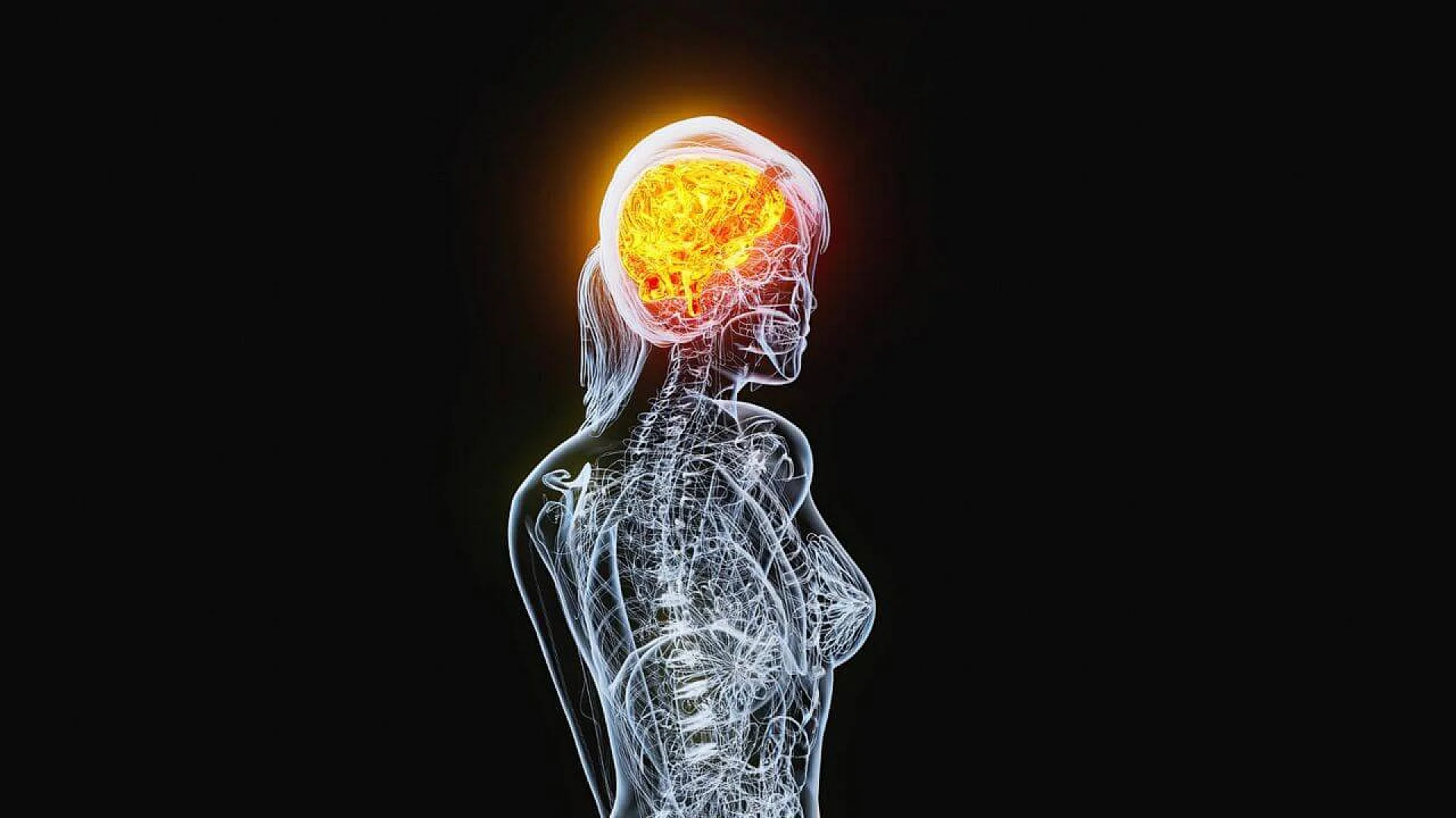
The human brain is not a fixed statue, it is a living organ that breathes with physiology. A new within-subject MRI study finds that the regular rise and fall of menstrual hormones are linked to short-term shifts in brain structure, spanning white matter microstructure, cortical thickness, and fluid versus tissue volume. The research suggests that hormone rhythms that repeat hundreds of times across a lifetime can measurably reshape brain architecture on the timescale of days.
The problem: why this matters and what we did not know
Researchers have long known that sex steroids and pituitary gonadotropins influence brain function, cellular plasticity, and behavior. What has been harder to establish is whether the daily and weekly hormone fluctuations of a normal menstrual cycle also cause detectable, repeatable changes in the brain’s anatomical highways (white matter tracts) and nodes (gray matter regions). Previous imaging studies were limited by small samples, coarse diffusion metrics that are confounded by fiber crossings, and reliance on phase estimates rather than direct hormone assays. The new study set out to close these gaps by combining direct hormone measurement, advanced diffusion imaging, and high-resolution cortical thickness mapping in a robust, within-subject design.
The approach, in plain language
The team recruited 30 healthy, naturally cycling women, each scanned and blood sampled three times timed to estimated menses, ovulation, and mid-luteal phases. Hormone assays measured 17β-estradiol and progesterone by highly sensitive LC-MS/MS, and luteinizing hormone (LH) and follicle-stimulating hormone (FSH) by automated immunoassay. MRI included a T1 MPRAGE anatomical scan for cortical thickness and a multidimensional diffusion protocol (q-tensor imaging, QTI) that provides subvoxel estimates of diffusion properties. Rather than rely on conventional diffusion tensor metrics that can be confounded by crossing fibers, the scientists used multidimensional diffusion parameters such as Diso (an MD-like index), μFA (micro-fractional anisotropy, robust to fiber crossings), and other size, shape, and orientation metrics. Statistical inference used hierarchical Bayesian regression models that linked within-participant hormone fluctuations to within-participant brain changes.
To use an analogy, conventional diffusion imaging is like measuring traffic speed at a single intersection and assuming all roads meet neatly. Multidimensional diffusion is more like using a 3D traffic camera that separates lanes and distinguishes intersecting flows, so you can detect subtle changes in particular routes even when they cross. This makes the new measures more sensitive and specific to microstructural tissue changes.
The breakthrough discovery
A quick summary of the headline findings
- Across the whole brain, higher 17β-estradiol and higher LH levels were associated with greater diffusion anisotropy measured by μFA. This suggests that these ovulatory hormones track increases in directional diffusion consistent with changes in white matter microstructure.
- FSH concentrations were positively associated with mean cortical thickness at the whole-brain level. In other words, pituitary gonadotropin dynamics tracked changes in gray matter thickness.
- Progesterone showed opposing volumetric effects: it was credibly associated with increased tissue volume and decreased cerebrospinal fluid (CSF) volume while total brain volume remained stable. That pattern points to hormone-linked shifts in water distribution and tissue properties.
White matter details: MD (Diso) and anisotropy (μFA)
At the whole-brain level, 17β-estradiol and LH were positively associated with anisotropy measures (μFA and D²anison), indicating more directional diffusion when those hormones are higher, consistent with changes in organizational tissue properties or water distribution along fibers. FSH and progesterone produced opposing effects on Diso, an index akin to mean diffusivity. Regionally, FSH decreases in Diso were observed in 17 white matter regions, while progesterone increases in Diso were found in seven overlapping tracts. The overlapping regions include limbic and visual system tracts, such as segments of the corpus callosum, fornix, optic radiation, and posterior thalamic radiation. In plain terms, pituitary and ovarian hormones push mean diffusivity in opposite directions in some of the same tracts.
Gray matter details: cortical thickness
At the whole-brain level, FSH was the hormone most clearly associated with cortical thickness. Regionally, both FSH and progesterone related to CT in several shared cortical areas, including fusiform gyrus, parahippocampal gyrus, lingual gyrus, isthmus cingulate, lateral orbitofrontal cortex, and pericalcarine cortex, but often with opposite directions. Progesterone tended to show negative CT relationships in several inferior and limbic regions while sometimes showing positive associations elsewhere, revealing a regionally heterogeneous pattern.
Brain volume and water dynamics
Although total brain volume did not shift credibly across hormone levels, progesterone associated with increased tissue volume and decreased CSF volume. This suggests hormone-linked redistribution of water and tissue compartments rather than gross brain size change, consistent with rapid, reversible morphological changes. Such effects might reflect altered extracellular water, cellular swelling, vascular changes, or combinations thereof.
Why it matters: implications for neuroscience, behavior, and clinics
- A dynamic brain tied to normal physiology. The study shows that normal menstrual hormone rhythms are associated with repeatable, measurable structural brain changes on timescales of days to weeks. That reframes the cycling brain as an organ that routinely remodels, not only during dramatic life events such as puberty or pregnancy, but month by month.
- Functional consequences are plausible. Structural changes that affect white matter anisotropy and cortical thickness may alter network communication and functional activation. This offers a plausible mechanism linking hormonal cycles to variation in mood, cognition, and sensory processing reported by many people who menstruate. The paper does not report behavioral correlations, but it motivates follow-up work to connect structure, function, and symptoms.
- Pituitary gonadotropins deserve attention. FSH, often overlooked in brain studies compared to estradiol and progesterone, showed robust associations with both white and gray matter architecture. That raises questions about direct or indirect actions of FSH on brain tissue and highlights the need to study the broader hormonal milieu.
- Clinical relevance for injury and treatment. If hormones modulate water dynamics and tissue microstructure, they might influence recovery after brain injury or alter responses to neurological disease. Understanding these effects could inform sex-specific clinical strategies and timing of interventions.
Caveats, limitations, and next steps
No single study answers everything. The scientists highlight several important limitations to interpret the findings responsibly:
- Sparse sampling. Each participant was sampled at three time points. That captures large cycle-related hormone swings but misses day-to-day microdynamics. Dense daily sampling would refine the timing and causal inference.
- Ovulation timing. Scheduling relied on at-home ovulation tests and cycle prediction, which can be imprecise. Some “ovulation” sessions may not reflect the exact biological ovulation day for every participant.
- Correlation, not causation. The results show co-fluctuation, not proof that hormones cause the brain changes. Experimental hormone suppression or replacement studies are needed to establish causality.
- Sample constraints. Participants were healthy adults aged 18 to 29, nulliparous, and not on recent hormonal contraception. Findings may not generalize across age, reproductive history, or clinical populations.
- Multiple models and conservatism. The analysis involved many Bayesian models. The team used ROPE thresholds derived from null simulations to reduce false positives, but replication in larger samples or consortia is important.
Conclusion
This study provides clear evidence that circulating HPG-axis hormones co-fluctuate with measurable changes in both white matter microstructure and cortical thickness across the menstrual cycle. The effects are widespread, occurring in classic hormone-receptor dense regions and across frontal, parietal, temporal, and occipital areas. Taken together, the findings argue that the brain of a person who menstruates is dynamic on a routine timescale, shaped in part by normal endocrine rhythms. Future dense-sampling, multimodal, and interventional work will be required to map cause, mechanism, and behavioral consequences, but the present study offers a precise, rigorously measured first view of a cyclical, remodeling human brain.
The research was published in Human Brain Mapping on July 19, 2024.
This article has been fact checked for accuracy, with information verified against reputable sources. Learn more about us and our editorial process.
Last reviewed on .
Article history
- Latest version
Reference(s)
- Rizor, Elizabeth J.., et al. “Menstrual cycle-driven hormone concentrations co-fluctuate with white and gray matter architecture changes across the whole brain.” Human Brain Mapping, vol. 45, no. 11, 19 July 2024, doi: 10.1002/hbm.26785. <https://onlinelibrary.wiley.com/doi/10.1002/hbm.26785>.
Cite this page:
- Posted by Dayyal Dungrela