a
b
c
d
e
f
g
h
i
j
k
l
m
n
o
p
q
r
s
t
u
v
w
x
y
z
#
Zoology
An Introduction to Protozoa
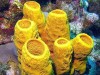
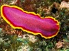
An Introduction to Sponges (Porifera)
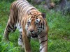
Animals Found in the Jungle: A Comprehensive Guide to Jungle Wildlife
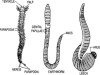
Annelida: Exploring the Diversity and Classification of Segmented Worms
Aurelia: Gastro Vascular System and Nutrition
Autonomous Nervous System, Sympathetic Nervous System and Parasympathetic Nervous System
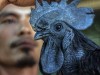
Ayam Cemani: Unveiling the Mystique of this Exotic Chicken Breed
Biology of Annelids
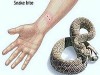
Biting Mechanism of Snakes: How Snakes Inject Venom Efficiently
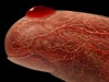
Blood Drop
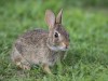
Breathing and Mechanism of Gas Exchange Respiration in Rabbit
Can Freshwater Fish Survive in Saltwater? Exploring the Science Behind Adaptation
Canal System in Sycon (Sponge)