Medically reviewed and approved by a board-certified member

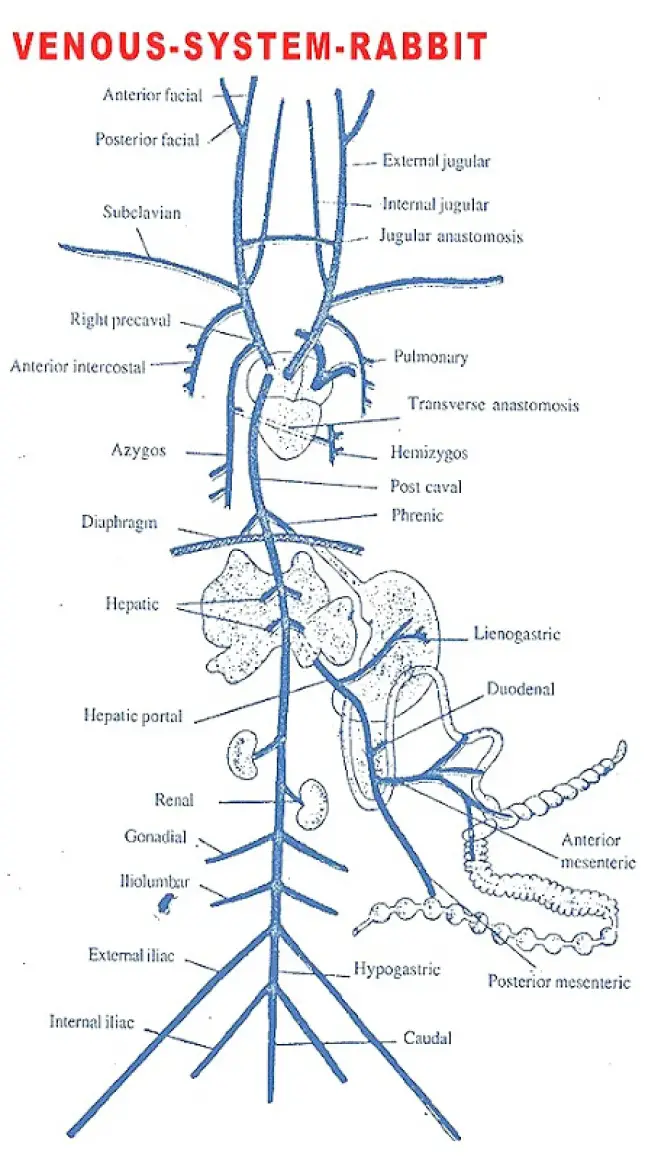
The blood from various parts of the body is collected by different types of veins which constitute venous system. Coronary veins collect the venous blood from the wall of heart into the left precaval vein.
The blood from the anterior and posterior ends of the body is mainly collected by two precavals or superior vena cavae and one post caval or inferior vena cava respectively.
Each precaval is formed by the union of three veins namely
- External jugular vein
- Internal jugular vein
- Sub-clavian vein
The external jugular vein brings the blood from tongue, jaws and muscles of the head.
- The external jugular is connected with its fellow of the opposite side by a transverse or jugular anastomosis that anastomoses in the neck.
- The internal jugular vein receives internal carotid vein collecting blood from the brain and neck.
- Then it runs down the neck along the side of the trachea and opening into external jugular vein opposite to the sub-clavian.
- The sub-clavian vein is a large vein on each side of the body. It collects blood from the shoulders and forelimbs.
- The two precavals open into the right auricle.
Each precaval vein also receives two smaller veins on either side namely,
i. Anterior intercostal vein and
ii. Azygos cardinal vein.
- Anterior intercostal vein collects blood from the anterior inter-costal spacfes and joins the precaval.
- This vein is formed by the union of four or five smaller veins.
- Azygos cardinal vein is formed by the union of small veins and opens into the right precaval vein.
- On the left side of the animal Azygos vein is absent, but few small veins form a hemizygos vein.
- Hemizygos vein joins the right azygos vein through a transverse Anastomosis.
- The post-caval vein or Inferior vena cava is a large median vein formed in the posterior part of the abdominal cavity by the union of several veins.
- Internal iliacs or hypogastric of the two sides unite along the middle line together with a median caudal vein from the tail to form a common hypogastric vein.
- The internal iliacs bring the blood from the back of the thighs.
- The common hypogastric vein receives a large external iliac vein
on either side. Each external iliac vein is formed by
- a femoral vein bringing blood from the inner or preaxial side of the thigh and
- a posterior epigastric vein collecting blood from ventral wall of the abdomen.
- A small vesicular vein also brings the blood from urinary bladder into the common hypogastric vein.
- The post caval vein runs anteriorly and it receives number of veins from different organs on its way to the right auricle.
- In front of external iliac, the post caval vein receives a pair of large ilio-lumbar veins which bring blood from the hinder part of the walls of abdomen.
- Anteriorto the ilio-lumbar a pair of gonadial veins also open into the post caval, spermatic veins from testes of male and ovarian veins from ovaries of female.
- In front of the gonadial veins the post caval receives two large renal veins collecting blood from the two supra renals and kidneys.
- The right renal vein is slightly anterior than the left renal vein.
- The renal portal system is absent in the rabbit. Post caval vein further runs anteriorly through the liver from which it receives blood by several hepatic veins.
- Hepatic portal system is present in rabbit and it consists of a hepatic portal vein ventral to the post caval vein.
- It enters into liver, divides into number of branches to supply to various lobes of the liver.
The hepatic portal vein is formed by several veins like
i. A lienogastric vein that brings blood from the stomach and spleen
ii. A duodenal vein collecting blood from duodenum and pancreas
iii. An anterior mesenteric vein bringing blood from the caecum, colon and part of the rectum, and
iv. Posterior mesenteric vein that collects blood from lower part of the rectum.
- The blood from the lungs is collected by a pair of large pulmonary veins.
- The two pulmonary veins open by a common aperture into the left auricle.
- The pulmonary veins bring oxygenated blood from lungs into left auricle.
Tags:
End of the article