COMPARATIVE ANATOMY: SKULL OF FISH, FROG, LIZARD, BIRD AND RABBIT
The hard parts of the animal body are collectively known as the skeletal systems or simply skeleton. The vertebrates possess the hard parts inside the body. It is known as an endoskeleton. The endoskeletal structures are formed with cartilages and bones which are the living tissues. The endoskeleton has been divided into: The hard parts of the animal body are collectively known as skeletal system or simply skeleton. The vertebrates possess the hard parts inside the body. It is known as an endoskeleton.
The endoskeletal structures are formed with cartilages and bones which are the living tissues. The endoskeleton has been divided into:
- Axial skeleton - includes the skull and vertebral column.
- Appendicular skeleton - includes the girdles and limb bones.
Also read COMPARATIVE ANATOMY: Respiration in Birds, Reptiles, and Mammals
The skull develops in the head of the animal body. The skull includes two major parts - 'Cranium' enclosing the brain and the organs of special sense and Visceral arches' which form the jaws and framework of the pharyngeal wall.
The cranium is developed from the mesodermal cells soon after the appearance of the brain. It is also known as a brainbox. Cranium includes three pairs of capsules for smell, sight, and hearing. These are known as olfactory, optic, and auditory capsules respectively. The cartilaginous cranium is called chondrocranium and the bony cranium is called dermato cranium.
The visceral arches develop around the anterior (Pharyngeal) part of the embryonic gut from the cells of neural crests. Mostly seven visceral arches are present. The first one is the largest and highly modified - 'Mandibular arch. It has dorsal & ventral halves. Each side of the dorsal half is termed the palato -pterygoid Quadrate Cartilage. It bears teeth and forms the upper jaw. The ventral half of the mandibular arch is called Meckel's cartilage. It also bears the teeth and forms the lower jaw. The wide gap between the two jaws is the mouth. The two jaws articulate their hind ends by hinge joints which enable the mouth to open & close. The second arch is the hyoid arch and the remaining five arches are termed bronchial arches. The visceral arches are collectively known as the splanchnic cranium. The upper jaw and lower jaw are known as Maxilla and Mandible respectively:
| S. No. | Skull of Scoliodon (Shark) | Skull of Rana (Frog) | Skull of Calotes (Garden Lizard) | Skull of Columba (Pigeon) | Skull of Oryctolagus (Rabbit) |
|---|---|---|---|---|---|
| 1. | Skull is formed with cartilage tissues. | Skull is formed mostly with bony tissues (but tadpole skull is cartilaginous) | Skull is formed mostly with bony tissues. | Skull is formed mostly with bony tissue. | Skull is formed with mostly bony tissue. |
| 2. | It consists of the cranium, sense capsules, and visceral arches. | It consists of a cranium, sense capsules, jaws, and hyoid apparatus. | It consists of a cranium, sense capsules, jaws, and hyoid apparatus. | Same as in calotes. | Same as in calotes. |
| 3. | It is the axial portion of the skull. It is more or less a violin box open in front and behind with an arched roof and flattened floor. It is divided into occipital, auditory, orbital, and ethmoidal regions. | It forms the middle hollow part of the skull. It is divided into auditory, olfactory, and occipital regions. | It forms the median hollow part of the skull. It is divided into occipital, auditory, orbital, olfactory, and optic regions. | It forms the posterior median hollow part of the skull. It is divided into occipital, auditory, optic orbital, and olfactory regions. | It forms the middle hollow part of the skull. It is divided into occipital auditory, optic orbital & olfactory regions. |
| 4. | Foramen magnum is posteriorly present. | Same. | Same. | Same. | Same. |
| 5. | Beneath the foramen magnum, a deep concavity is present. On either side of this concavity is a prominence - occipital condyle articulates with the first vertebra, occipital crest is formed. Dicondylic skull. | Beneath the foramen magnum, there are two occipital condyles. On either side of the foramen, magnum dorsolaterally exoccipital bones are present. Dicondylic skull | Beneath the foramen magnum a single occipital condyle is present.suupraoccipitai, exo occipitals,& basi occipital bones are also present in the occipital region. Monocondylic skull. | Beneath the foramen magnum single occipital condyle is present. Supra occipital, Exocci pitals & basioccipital bones are also present. Monocondylic skull. | Beneath the foramen magnum two occipital condyles with paroccipital process are present. Supraoccipital, exo-ccipitai, & basio-ccipital bones are also present. Dicondylic skull. |
| 6. | The auditory region has a mid-dorsal depression - parietal fossa. It contains two pairs of apertures. Anteriorly smaller openings of endolymphatic ducts and posteriorly larger openings of perilymphatic spaces are present. | — | — | — | — |
| 7. | Auditory capsules lie on the poster lateral sides of the cranium. Which enclose & protect the ears. Post orbital groove is present on the ventral side | Auditory capsules enclose the internal ear. Its roof is formed by pro-otic bone, fenestra ovalis, stapedial plate and columella auris are present. | Each auditory capsule is formed by a small, single vertical prootic bone that is lying outside the supraoccipital. Epiotic & opisthotic are not differentiated. | Each auditory capsule is formed largely by the prootic bone. Fenestra ovalis, fenestra rotun da, columella auris, stapes are also present. | Each auditory capsule in the adult animal consists only periotic. Flask-like Tympanic bulla bone is significant. |
| 8. | — | Dorsally the cranium is formed, by the frontoparietal, ventrally by parasphenoid, and laterally by sphenethmoid bones. | The dorsal part of the cranium is formed by parietals, frontals interparietal foramen, and ventrally by basisphenoid, parasphenoid bones. | The dorsal part of the cranium is formed by Parietals, frontals a rostrum, alisphenoids; ventrally basisphenoid, bitemporal bones. | The cranium is formed dorsally by 'Parietals, frontals, interparietal, and ventrally by basisphenoids, presphenoid bones along with alisphenoids and orbit sphenoids. The cranial cavity is closed in front by a narrow vertical bone cribriform plate. |
| 9. | Each orbit lies on the sides of the middle part of the cranium. It is bordered by a dorsal super orbital ridge, anterior preorbital process, posterior postorbital process, and ventrally by an infraorbital ridge. The orbital region has a large oral cavity anterior fontanelle. | On either side of the cranium is a large gap - orbit which lodges the eye. | In the middle of the cranium laterally two orbits are present. Each orbit is bounded by prefrontal supraorbital, lacrimal, post-frontal, and jugal bones. The jugal bone forms the ventral border of the orbit. Supratemporal arch is present. | The two orbits are very large cavities present in front of the cranium. Each orbit is bounded dorsally by frontal, anterodorsally by lacrimal, and posteriorly by the zygomatic process. Orbit is incomplete on the ventral side. The two orbits are separated by an interorbital septum. | These are two orbits that are large sockets present on the sides of a frontal segment of the cranium. The orbit is bounded dorsally by frontal, anteriorly by the maxilla and lacrimal, posteriorly by squamosal and alisphenoid, and externally by the zygomatic arch. |
| 10. | The olfactory capsules lie at the anterior side of the cranium. Each capsule possesses a short sic at the ethmopalatine ridge. | The olfactory capsules are separated, from each other by mesethmoid. Each capsule is formed by a large triangular nasal on the dorsal side and a smaller triradiate vomer on the ventral side vomers possess vomerine teeth. | Each olfactory capsule is formed by three bones Nasal, septo-maxillary, and vomer. | Each olfactory capsule is formed by two bones - Nasal and vomer. Nasals fuse with frontals and form into super and inferior processes. | Each olfactory capsule is bounded dorsally by long nasal bone and laterally by jawbones. The two capsules are separated by mesethmoid bone. The lower end of the mesethmoid fits into a vomer bone. The vomer is formed by the fusion of a pair of bones. |
| 11. | The ethmoidal region tapers anteriorly. It consists of a basal slender barventro-median rostral cartilage and a pair of similar barsdorso - lateral rostral cartilages arisen from the roof of the olfactory capsules. | Absent. | Absent. | Absent. | Absent. |
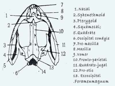
| 12. | Scoliodon has seven visceral arches which are cartilaginous. The first arch forms the jaws and it is catted the Mandibular arch the second one is the hyoid arch the remaining five arches are called branchial arches. | Branchial arches are absent. There are upper and lower jaws to support the borders of the mouth. The upper jaw is formed by the union of two similar halves. Each half is formed by the Pre-maxilla, maxilla, and quadratojugal. The inner set of the jaw has palatine, pterygoid and squamosal bones. The lower consists of two halves and unite anteriorly by mentomeckelian cartilage. Each half consists of the dentary and angio-splenial bones. Just in front of the articular fact, a small coronary process is present. The upper jaw alone has teeth. | Branchial arches are absent. | Branchial arches are absent. | Branchial arches absent. These are upper and lower jaws. Each half of the upper jaw is formed by premaxilla, maxilla jugular, palatine, pterygoid and squamosal. |
| 13. | The mandibular arch consists of two halves. Each half of this arch possesses an upper paletopterygo quadrate cartilage and a lower Meckel's cartilage. The paletopterygo Quadrate gives off anteriorly palatine. The two sides of it from the upper jaw with teeth. The two Meckel's cartilages united anteromedially by ligament from the lower jaw with teeth. | — | These are upper and lower jaws. Each half of the upper jaw consists of an outer set of bones - premaxilla, maxilla, jugal, and quadrate and the inner set includes pterygoid, palatine, transpalatine, epipterygoid, and squamosal. Each half of the lower jaw consists of six bones -dentary, angular, supra angular, articular, splenial, and coronoid. Both the jaws possess teeth. | These are upper and lower jaws. Each half of the upper jaw is formed by premaxilla, maxilla, quadra tojugal, and jugal bones. The inner arcade of the upper jaw forms the roof of the buccopharyngeal cavity which consists of palatine, pterygoid, and quadrate. Each half of the lower jaw is formed by articular, angular supra angular, dentary, and splenial. Both the jaws are lacking teeth. | The lower jaw also consists of two halves. Each half is formed by a single, large dentary bone. The posterior of the dentary possess condylar, coronoid and angular process. Both the jaws possess the codent type of teeth which are having different (Heterodont teeth in mammals) shapes. Diastema is present in both jaws because of the absence of canines. |
| 14. | Hyostylic jaw suspension. | Autostylic jaw suspension. | Autostylic jaw suspension. | Autostylic jaw suspension. | Craniostylic jaw suspension. |