Medically reviewed and approved by a board-certified member
- Contraction is the main property of muscle.
- The muscle receives stimuli through the motor nerve.
- The teledendrites of last neurons of motor nerve, which innervate in the muscle are called "neuromotor end plates".
- When the nerve impulse passes to the neuromotor end plates, "acetyl choline" is released.
- Acetyl choline stimulates the muscle to contract.
- After the muscle starts contraction acetyl Choline is destroyed immediately by "choline esterase".
- The muscle contracts totally or does not contract.
- The strength of contraction depends upon the intensity of stimulus.
- Muscle contracts due to nerve impulse.
- But for stimulation to contract, the muscle fibre always requires a specific minimum strength of nerve impulse or stimulus.
- This is called "Threshold stimulus".
- The single isolated contraction of muscle fiber is called "Muscle twitch'.
- When the muscle fiber is stimulated by rapid nerve impulses, the muscle fiber remains in a state of contraction so long as the stimulation continuous.
- Such a continued state of contraction is called "Tetanus".
- The muscle fiber always contracts with maximum force irrespective of the intensity of stimulus.
- The force of contraction does not increase even though the intensity of the stimulus is increased.
- This is known as "all-or-none law". But muscle does not contract when the stimulus is below the threshold stimulus.
- The muscles which contract to produce opposite movements at the same joint are called "Antagonistic muscles".
- When a muscle contracts, its antagonistic muscle must relax to allow the movement.
- The biceps is a flexor muscle which, folds the elbow joint and triceps is an extensor, the muscle which extends the elbow joint.
- Also read structure of muscle
MECHANISM OF MUSCLE CONTRACTION
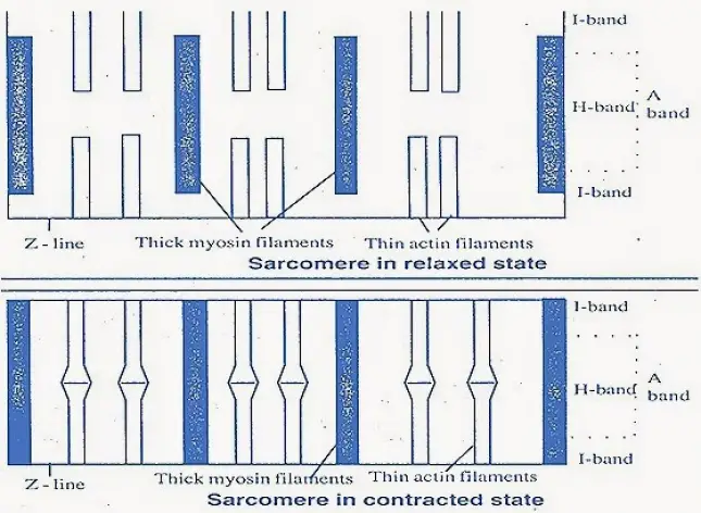
- When the muscle is in relaxed state, I- bands and H - bands are longer and A - bands or thick bands are shorter, when the muscle is in contracted state, I - bands and H - bands are smaller but A -bands or thick bands are larger.
- This change occurs in the entire length of sarcomere.
- The muscle contraction was explained by sliding filament theory.
SLIDING FILAMENT THEORY
- It was proposed by H.E. Huxley and A.F. Huxley [Jean Hanson].
- According to them muscle becomes shorter and shorter, the thick and thin filaments slide past each other.
- But the length of filaments is not altered. As the thin filaments slide past the thick filaments, they thin filaments] meet in the centre of sarcomere.
- Therefore the TV-band never disappears.
- But the l-band shortens and disappears.
- The actual sites of contraction are the cross-bridges, which are formed when the two filaments overlap. During relaxation, the cross bridges break.
- Hence the thin actin filaments move away from 'A' band. Then the l-band reappears.
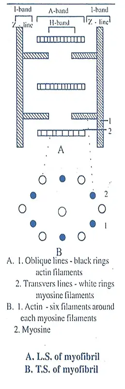
- Each thick or myosin filament is surrounded by six actin filaments in hexagonal form.
- Each cross bridge is attached with actin filament.
- At this place of contact, actin and myosin filaments are attached with each other forming acto-myosin which initiates contraction, i.e. the actinfilaments slide past the thick myosin filaments.
- Each cross bridge is formed of one ATP molecule. For every second cross bridge disappears and appears for 50 to 100 times
RATCHET MECHANISM
- During contraction and relaxation, the cross bridges operate in a cyclic manner alternately attaching and detaching from thin actin filaments.
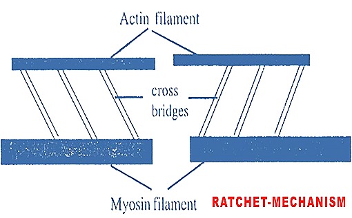
- This is called ratchet mechanism. The bridges act like hooks or levers.
- A number of mitochondria are present in between the filaments which provide ATP neccessary for muscle contraction.
CHEMICAL EVENTS
During muscle contraction, there occurs many chemical events. These are first worked out by "Szent Gyorgi."
i. Whenever the acetyl choline is released on the muscle, the Ca++ are released from endoplasmic reticulum [sarcoplasmic reticulum].
Acetyl choline + E.R----------------> Ca++
ii. The enzyme myosin A.T.P. ase is active in presence of Ca++, and Mg++. Which are also essential for muscle contraction. The A.T.P. ase breaks ATP into ADP, P [phosphate] and energy in presence of Ca++ and Mg++ by hydrolysis.
ATP + H2O -----------> ADP + P + Energy
iii. The thick myosin filaments now binds the thin actin filaments by using energy. Hence "Actomyosin" is formed.
Actin + Myosin ------------------> Actomyosin
iv. For relaxation also, muscle requires energy. Hence ADP is immediately changed to ATP by creatine phosphate
ADP + CP------------> ATP + C [creatine]
Now again the ATP breaks into ADP to release energy. Such energy is used to break down the actomyosin. Hence thin filaments move to the normal position. Thus the l-band reappears.
Actomyosin + Energy.. --------------> Actin + Myosin
vi. The creatine produced is converted into creatine phosphate by ATP.
Creatine + ATP --------->ADP + CP
[creatine phosphate]
[creatine phosphate]
vii. The ATP is synthesized by the oxidation of carbohydrates [glucose].
C6H12O6 + O2 --------------> CO2 + H2O + ATP
viii. The muscle stores glucose in the form of glycogen. During strenuous exercise, the muscle does not get sufficient oxygen. Hence muscle respires by anaerobic glycolysis. Glycolysis in the muscle was explained by "Cor/and Corf. They proposed the glycolysis in muscle and liver as Cori's cycle. The muscle glycogen is broken down into lactic acid and energy. The energy is used for the synthesis of CP. The lactic acid is released into blood, which is circulated to the liver. In liver 4/5 of lactic acid is converted into glucose and 1/5 is oxidised to CO2 and H2O. The glucose is transported to the muscle through blood. In muscle glucose is converted into glycogen. The steps in Cori's cycle are given below.
STRETCH REFLEX
- Muscle spindle is present parallel to muscle fibres of a muscle.
- In the middle part of muscle spindle, a bundle of small muscle fibers present.
- They are non-contractile.
- The two ends of muscle spindle contain spinal nerve endings and are contractile.
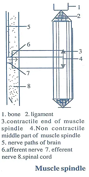
- The two contractile ends of muscle spindle are supplied with afferent and efferent nerves from central nervous system.
- As and when muscles expand, the nerve impulses are transmitted from muscle endings to central nervous system through afferent nerves.
- The muscles receive responses accordingly from CNS through efferent nerves.
- As a result, the.two endings of muscle spindle undergo contraction.
- This is called 'stretch reflex'.
Tags:
End of the article