Medically reviewed and approved by a board-certified member
Zoology
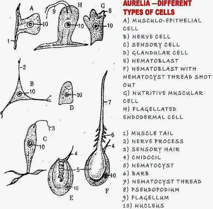
HISTOLOGY OF AURELIA (JELLYFISH CELLS)
By BS MediaTwitter Profile | Updated: Tuesday, 18 April 2017 20:35 UTC
Jellyfish or Aurelia is a marine sedentary coelenterate animal.
The body wall has two layers. The outer layer is ectoderm. The inner layer is called gastrodermis or endoderm. In between these two layers jelly like mesoglea is present.
Ectoderm:
It covers the body and gives protection. It is a single layer. It is also useful in reproduction. In this layer 6 types of cells are present.
1. Epithelio-muscular cells: In the ectoderm of obelia these cells are more in number. They form the covering on the body. The outer part of the cell is broad. The inner side is narrow. The broader ends of nearby cells unite and a layer is formed. From the narrow end two muscular tails will arise. In each tail two myonemes are present. They can undergo contraction. These muscle tails are arranged longitudinally, they form longitudinal muscles. When these muscles contract animal becomes short. The broader end of the cell will show small granules. They secrete cuticle.
2. Interstitial cells: These are present in between the musculo-epithellial cells. They are small and round. They are totipotent cells. They can change into any other kind of ectodermal cell. They are also called formative cells. They produce buds and sex cells.
3. Nerve cells: They are also called protoneurons. They are present on the base of the ectoderm. Each cell gives rise to nerve fibers. All these nerves will unite to form a net work. This nerve network is more near the hypostome. Such a nervous system is called diffused nervous system. This nerve net is connected with sensory cells.
4. Sensory cells: These cells are long and narrow. They are more in tentacles and base. On the free end of each cell a flagellum or a process is present. On the inner side of the cell two fibers are present. They are connected with nerve network. The nerve network and sensory cells are combindly called neuro motor system.
5. Glandular cells: In the base and hypostome these gland cells are more in number. They contain granules. They produce mucous.
6. Cnidoblast (or) Nematoblast : The nematocytes of aurelia are 1. Atrichousisorhiza 2. Holotrichousisorhiza 3. Heterotrichousmicrobasiceuryteles. These are also called stinging cells. These cells are present in the entire ectoderm. They are more in the tentacles. These are formed by the modification of interstitial cells. When they are completely formed they are distributed in the entire ectoderm.
Structure of heterotrichous microbasic euryteles: In each cnidoblast a central bag like structure is present, it is called nematocyst. It is filled with a fluid called hypnotoxin. In this nematocyst a long thread is coiled. The nematocyst is closed by operculum. Near the operculum a cnidocil is present.
Around this nematocyst cytoplasm is present. In this cytoplasm the nucleus is pushed to one side. In the cytoplasm contractile fibers are present. They are also large fibres. This fibre will not allow the nematocyst to go out of the cnidoblast.
On the base of the tube spines will be present. The tube opens at the tip.
The cnidoblasts are useful for self defense, food collection and to attack on other organisms, if the prey comes in contact with cnidocil, the nematocyst is discharged. A long thread which is coiled inside will become straight, it pierces into the preys body. The prey becomes paralysed. Since the thread is cut off and remains in the body of the prey the nematocyst functions only once.
Mesoglea:
In Aurelia in between ectoderm and gastodermis a non cellular, jelly like mesoglea is present. With the help of this mesoglea the two layers are united. This layer is produced by ectoderm and gastodermis cells. This layer gives support to the two layers.
Cells of Gastodermis:
This layer is present inside the body, it contains five types of cells. This layer will digest the food.
1. Nutritive muscle cells: These are also called nutritive cells. They look like epidermal muscle epithelial cells. The narrow part shows contractile fibres. These are arranged transversely. They work like circular muscles. The muscles are mainly present near mouth. They work as sphincters. They can close the mouth. The free end of the cells is facing coelenteron. On this free end flagella or pseudopodia will be present. The pseudopodial cells will catch half digested food and digest the food. This is called intracellular digestion.
2. Glandular cells: These cells are small in size. They may contain one or two flagella. The cells near the mouth will produce mucilage. Cells in the inner side will produce digestive juices.
3. Interstitial cells: In the endoderm in between two nutritive cells these cells are present. They are formative cells.
4. Nerve cells: They look like nerve cells of ectoderm. Their number is less they may not form a net work.
5. Sensory cells: They look like ectodermal cells Their function is not clearly known. They are few in number. They are distributed among nutritive cells
6. Cnidoblasts : These are confined to gastric filaments.
7. Germ cells: Interstitial cells will develop into ova or sperms.
End of the article