Medically reviewed and approved by a board-certified member
Cell Biology
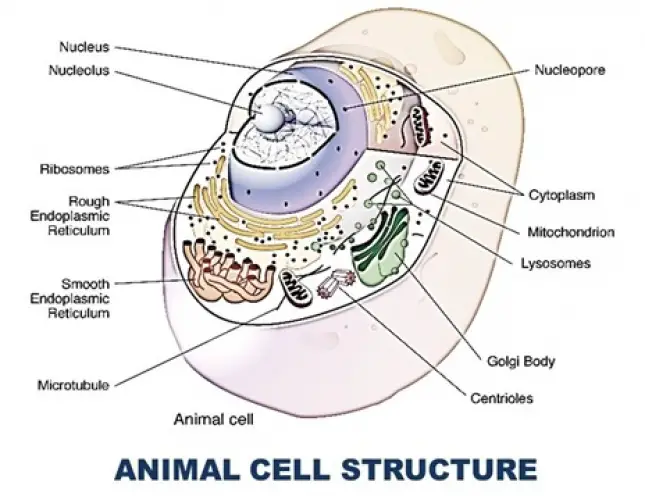
AN ANIMAL CELL STRUCTURE
By BS MediaTwitter Profile | Updated: Tuesday, 06 June 2017 17:41 UTC
Cell is a fundamental, structural and functional unit of living organism. The science which deals with cells and their organelles is called cell-biology. The term cell was first used by Robert Hooke in 1665. He described the cell first as cella which means hollow space. Robert Hooke observed cells in the section of cork. In 1831 Robert Brown observed nucleus in plant cells. In 1858 Rudolf Virchow stated that new cells arise from pre-existing cells.
"Omnis cellulae cellula. Schilden, German Botanist in 1938 described cell theory with regard to plant cell. T. Schwann German Zoologist in 1939 described cell theory with regard to animal cells. Cell theory denotes that "Cell is the structural and functional unit of life."
SHAPE OF CELL:
The shape of the cells is variable. The cells of unicellular forms, leucocytes and-bacteria, exhibit a number of shapes and those of multicellular organisms exhibit still further variation. Their shapes may be rounded, cylindrical, irregular, triangular and tubular.
SIZE OF CELLS:
Size is extremely variable, measuring from one micron to 175 mm. The ostrich egg cell is 176 mm. in diameter, thus visible to the naked eye. The nerve cell found in mammals may reach a length of 3 or 3.5 feet. Smaller cells are those of the Pleuropneumonia like organisms.
Plasma Membrane:
A porous membrane surrounds the cytoplasm called plasma membrane. Electron microscopic studies reveal that the plasma membrane is composed of outer, inner protein layers and in between them double layered lipids are present Robertson called plasma membrane unit membrane.
The main function of plasma membrane is to regulate the entry and exist of substances.
Cytoplasm:
The part of protoplasm outside the nucleus is known as cytoplasm. It is distinguished as an outer non-granular thick ectoplasm and inner granular thin endoplasm. In the cytoplasm many organelles are present.
Cell Organells:
In the cytoplasm many cell organelles are present.
1. Centrosome:
It is the center of the cell discovered by van Benden in 1887. It is found near the nucleus and includes a specialised portion of cytoplasm, called centirosome. Its matrix is called as kinoplasm, in which two centrioles are embedded. Each centriole consists of nine fibrillar units and each fibrillar unit is found to contain three microtubules. The function of centrioles is to form the spindle at the time of cell division.
2. Endoplasmic Reticulum:
In the cytoplasm a network of tubules are present. It is called endoplasmic reticulum. This network of tubules will be two types.
i. Smooth endoplasmic reticulum:
On the surface of the tubules ribosomes are absent. Hence they are ce Sled smooth endoplasmic reticulum or agranular endoplasmic reticulum.
ii. Rough endoplasmic reticulum:
On the surface of the tubules ribosomes are present. It is rough endoplasmic reticulum. This is called granular endoplasmic reticulum.
Endoplasmic reticulum will connect plasma-membrane nucleus and other organells.
Functions:
- It gives strength to the cell and forms cytoskeleton.
- Granular endoplasmic reticulum will produce proteins.
- Agranular endoplasmic reticulum will produce lipids.
- It forms the work bench for many biochemical reactions in the cell.
3. Ribosomes:
It is a small particle present in the cytoplasm. They will be attached to the cell organelles and they are also freely distributed in the cytoplasm.
In a Eukaryotic cell 80s ribosomes are present. This ribosome is made by 2 sub units. They are 40s, 60s sub units.
Ribosome is made by proteins and RNA. Ribosome shows 150 to 200A° diameter. Ribosome combine with m RNA and produce proteins. A group of ribosomes with m RNA is called polysome.
4. Golgi Complex:
They are described by Golgi. They are also called dictyosomes, Bpochondria, and idosomes. The complex shows three types of structures,
a. Cisternae:
These are flat sacs. They are arranged one above the other. They are 150 V in length, 60 A° in thickness.
b. Vacuoles:
These are oval in shape. They are big.
c. Vesicles:
They are in the form of groups. All these structures totally called Golgi complex.
Functions:
They are more in secretory cells. Hence they are connected with secretory function. They store proteins and lipids. During cell division they produce cell, plate. During the formation of sperm they will form the acrosome of the sperm.
5. Mitochondria:
These are first described by Altamann as Bioplasts, in 1894. They were m i led as mitochondria by Benda in 1897.
They are filamentous or rod like structures. The mitochondria are covered by layers. Inner membrane is folded inside. Those folding's are called cristae. On these cristae oxysomes are present.
- In the central matrix of mitochondria respiratory enzymes are present. The take up Krebs cycle reactions.
- In the inner membrane of mitochondria electron transport enzymes are present.
- Mitochondria helps in the oxidation of the food material and liberates energy , Hence they are called power houses of cell.
- In the mitochondria a circular DNA is present. Hence mitochondria is also c. led semi autonomous body.
6. Lysosome:
These are described by De-Duve. Each lysosome is round in shape. It shows .4 to .8 microns in diameter. It is covered by lipoprotein layer. It contains hydrolytic enzymes. It is useful for intracellular digestion and autolysis of the cell.
Functions :
- Lysosome is helpful in the digestion of the food.
- At starvation lysosome will digest cell organelles.
- Lysosome can dissolve the cell. It is called suicide. Henc lysosomes are called suicidal bags of cells.
Inclusions of the Cytoplasm:
In the cytoplasm vacuoles and duetoplasmic bodies are pr-sent. In the young stages vacuoles are absent in the cytoplasm. When the cell is growing the .Cytoplasm vacuoles are formed. In a older cell big vacuole is present. It is filled with cell sap. A vacuole is covered by tonoplast. In the cell sap water, some excretory substances, some pigments, and other substances are present.
Duetoplasmic Bodies:
In the cytoplasm reserve food materials, excretory wastes a<\l secretory substances are stored. They are called duetoplasmic bodies.
Nucleus:
In a eukaryotic cell a definite nucleus is present. It is 5 to 25 microns in, size. It shows the following parts.
a) Nuclear Membrane:
Nucleus is covered by a nuclear membrane. It is made by 2 layers. In between the two layers perinuclear space is present. In the nuclear membrane small openings are present. Around each opening on the out side a small annulus is present.
Hammerling proved that nucleus is the seat of heredity through grafting experiments on Acetabularia.
b) Nucleoplasm:
Below the nuclear membrane nucleoplasm is present. In this glycoproteins, RNA and enzymes are present.
c) Chromatin Network:
In the nuclear spa many chromosomes are present. They are thin and filamentous. They are in the form of a network. On the chromosomes genes are present. They are units of heredity.
d) Nucleolus:
In the nucleoplasm one or two round structures are present They are called nucleoli. They contain proteins and RNA. They produce ribosomes.
Functions:
- It is the seat of life in the cell.
- It carries hereditary characters from one generation to another generation.
- It produces nucleic acids.
End of the article