How to Count Reticulocytes (Manual Method)
Reticulocytes
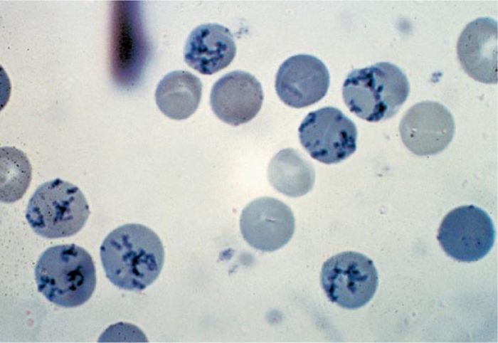
Reticulocytes are young or juvenile red cells released from the bone marrow into the bloodstream and that contain remnants of ribonucleic acid (RNA) and ribosomes but no nucleus. After staining with a supravital dye such as new methylene blue, RNA appears as blue precipitating granules or filaments within the red cells. Following supravital staining, any nonnucleated red cell containing 2 or more granules of bluestained material is considered as a reticulocyte (The College of American Pathology). Supravital staining refers to staining of cells in a living state before they are killed by fixation or drying or with passage of time. Reticulocyte count is performed by manual method.
PRINCIPLE
A few drops of blood (collected in EDTA) are incubated with new methylene blue solution which stains granules of RNA in red cells. A thin smear is prepared on a glass slide from the mixture and reticulocytes are counted under the microscope. Number of reticulocytes is expressed as a percentage of red cells.
REAGENT
New methylene blue solution is prepared as follows:
- New methylene blue: 1.0 gm
- Sodium citrate: 0.6 gm
- Sodium chloride: 0.7 gm
- Distilled water: 100 ml
Reagent should be kept stored in a refrigerator at 2-6°C and filtered before use.
Suitable alternatives to new methylene blue are brilliant cresyl blue and azure B.
SPECIMEN
Capillary blood or EDTA anticoagulated venous blood can be used.
METHOD
(1) Take 2-3 drops of filtered new methylene blue solution in a 12 × 75 mm test tube.
(2) Add equal amount of blood and mix well.
(3) Keep the mixture at room temperature or at 37°C for 15 minutes.
(4) After gentle mixing, place a small drop from the mixture on a glass slide, prepare a thin smear, and allow to dry in the air.
(5) Examine under the microscope using oil-immersion objective. Mature red cells stain pale green blue. Reticulocytes show deep blue precipitates of fine granules and filaments in the form of a network (reticulum). Most immature reticulocytes show a large amount of precipitated material in the form of a mass, while the most mature reticulocytes show only a few granules or strands. Any nonnucleated red cell is considered as a reticulocyte if it contains 2 or more blue-stained particles of ribosomal RNA.
(6) Count 1000 red cells and note the number of red cells that are reticulocytes. Counting error is minimized if size of the microscopic field is reduced. This is achieved by using a Miller ocular disk inserted in the eyepiece; it divides the field into two squares (one nine times larger in size than the other). Reticulocytes are counted in both the squares and the red cells are counted in the smaller square.
REPORTING THE RESULT
(1) Reticulocyte percentage: The most common method of reporting is reticulocyte percentage which is calculated from the following formula:
NRBC
Where NR is the Number of reticulocyte counted and NRBC is number of red blood cell counted.
Reference range is 0.5%-2.5% in adults and children. Reticulocyte count is higher in newborns.
(2) Absolute reticulocyte count = Reticulocyte percentage × Red cell count
Normal: 50,000 to 85,000/cmm
(3) Corrected reticulocyte count (Reticulocyte index)
Normal PCV
Corrected reticulocyte count > 2% indicates reticulocyte release appropriate for the degree of anemia. If < 2%, reticulocyte release is inappropriate.
(4) Reticulocyte maturation production index
Estimated maturation time in days
REFERENCE RANGES
- Reticulocyte percentage: 0.5 2.5%
- Absolute reticulocyte count: 50,000-85,000/cmm
References
- Pierre RV. Reticulocytes: Their usefulness and measurement in peripheral blood. Clin Lab Med 2002;22:63-79.
- The Expert Panel on Cytometry of the International Council for Standardization in Hematology: ICSH guidelines for reticulocyte counting by microscopy on supravitally stained preparations. World Health Organization. WHO/LBS/92.3 1992.
- Comment
- Posted by Dayyal Dg.